 Movie
Movie Controller


+ Open data

- Basic information
| Entry | Database: PDB / ID: 2r8f | ||||||
|---|---|---|---|---|---|---|---|
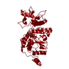
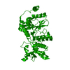
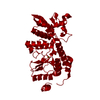
| Title | Crystal structure of H225A NSP2 and ATP-gS complex | ||||||
 Components Components | Non-structural RNA-binding protein 35 | ||||||
 Keywords Keywords | RNA BINDING PROTEIN / Rotavirus / NDP Kinase / non structural protein / NTPase / RNA-binding | ||||||
| Function / homology |  Function and homology information Function and homology informationnucleoside diphosphate kinase activity / viral genome replication / Hydrolases; Acting on acid anhydrides; Acting on acid anhydrides to facilitate cellular and subcellular movement / ribonucleoside triphosphate phosphatase activity / host cell cytoplasm / RNA binding / ATP binding / metal ion binding Similarity search - Function | ||||||
| Biological species |   Simian 11 rotavirus Simian 11 rotavirus | ||||||
| Method |  X-RAY DIFFRACTION / SYNCHROTRON / MOLECULAR REPLACEMENT / Resolution: 2.8 Å X-RAY DIFFRACTION / SYNCHROTRON / MOLECULAR REPLACEMENT / Resolution: 2.8 Å | ||||||
 Authors Authors | Kumar, M. / Prasad, B.V.V. | ||||||
 Citation Citation | Journal: J.Virol. / Year: 2007 Title: Crystallographic and Biochemical Analysis of Rotavirus NSP2 with Nucleotides Reveals a Nucleoside Diphosphate Kinase-Like Activity Authors: Kumar, M. / Jayaram, H. / Vasquez-Del Carpio, R. / Jiang, X. / Taraporewala, Z.F. / Jacobson, R.H. / Patton, J.T. / Prasad, B.V.V. #1: Journal: Nature / Year: 2002Title: Rotavirus protein involved in genome replication and packaging exhibits a HIT-like fold Authors: Jayaram, H. / Taraporewala, Z. / Patton, J.T. / Prasad, B.V.V. | ||||||
| History |
|
- Structure visualization
| Structure viewer | Molecule: MolmilJmol/JSmol |
|---|
- Downloads & links
-Download
| PDBx/mmCIF format | 2r8f.cif.gz | 78.9 KB | Display | PDBx/mmCIF format |
|---|---|---|---|---|
| PDB format | pdb2r8f.ent.gz | 58.8 KB | Display | PDB format |
| PDBx/mmJSON format | 2r8f.json.gz | Tree view | PDBx/mmJSON format | |
| Others |  Other downloads Other downloads |
-Validation report
| Arichive directory | https://data.pdbj.org/pub/pdb/validation_reports/r8/2r8fftp://data.pdbj.org/pub/pdb/validation_reports/r8/2r8f | HTTPS FTP |
|---|
-Related structure data
| Related structure data |  2r7cC  2r7jC  2r7pC  1l9vS S: Starting model for refinement C: citing same article ( |
|---|---|
| Similar structure data |


-Links
 PDBj
PDBj
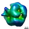
- Assembly
| Deposited unit | 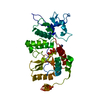
| ||||||||
|---|---|---|---|---|---|---|---|---|---|
| 1 | x 8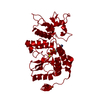
| ||||||||
| Unit cell |
|
-Components
| #1: Protein | Mass: 36551.191 Da / Num. of mol.: 1 / Mutation: H225A Source method: isolated from a genetically manipulated source Source: (gene. exp.) Simian 11 rotavirus (serotype 3 / strain SA11-Ramig)Species: Rotavirus A / Strain: Ramig / Gene: S8, SEGMENT 8 / Plasmid: pQE60 / Production host:  |
|---|---|
| #2: Chemical | ChemComp-PO4 /   Mass: 94.971 Da / Num. of mol.: 1 / Source method: obtained synthetically / Formula: PO4 Mass: 94.971 Da / Num. of mol.: 1 / Source method: obtained synthetically / Formula: PO4 |
| #3: Chemical | ChemComp-AGS /   Mass: 523.247 Da / Num. of mol.: 1 / Source method: obtained synthetically / Formula: C10H16N5O12P3S / Comment: ATP-gamma-S, energy-carrying molecule analogue*YM Mass: 523.247 Da / Num. of mol.: 1 / Source method: obtained synthetically / Formula: C10H16N5O12P3S / Comment: ATP-gamma-S, energy-carrying molecule analogue*YM |
| #4: Water | ChemComp-HOH /  Mass: 18.015 Da / Num. of mol.: 99 / Source method: isolated from a natural source / Formula: H2O Mass: 18.015 Da / Num. of mol.: 99 / Source method: isolated from a natural source / Formula: H2O |
-Experimental details
-Experiment
| Experiment | Method: X-RAY DIFFRACTION / Number of used crystals: 1 |
|---|
- Sample preparation
| Crystal | Density Matthews: 3.14 Å3/Da / Density % sol: 60.85 % |
|---|---|
| Crystal grow | Temperature: 298 K / Method: vapor diffusion, hanging drop / pH: 7.5 Details: 6-8% PEG 10000, 20mM Tris-HCl pH 7.5, 10-15mM nucleotide, VAPOR DIFFUSION, HANGING DROP, temperature 298K |
-Data collection
| Diffraction | Mean temperature: 100 K |
|---|---|
| Diffraction source | Source: SYNCHROTRON / Site: APS  / Beamline: 19-ID / Wavelength: 0.9790903 Å / Beamline: 19-ID / Wavelength: 0.9790903 Å |
| Detector | Type: ADSC QUANTUM 4 / Detector: CCD / Date: Dec 13, 2005 |
| Radiation | Protocol: SINGLE WAVELENGTH / Monochromatic (M) / Laue (L): M / Scattering type: x-ray |
| Radiation wavelength | Wavelength: 0.9790903 Å / Relative weight: 1 |
| Reflection | Resolution: 2.8→50 Å / Num. all: 11836 / Num. obs: 11744 / % possible obs: 99 % / Observed criterion σ(F): 3 / Observed criterion σ(I): 3 / Redundancy: 7.81 % / Rmerge(I) obs: 0.08 / Net I/σ(I): 14.9 |
| Reflection shell | Resolution: 2.8→2.9 Å / Redundancy: 7.97 % / Rmerge(I) obs: 0.281 / Mean I/σ(I) obs: 5.1 / Num. unique all: 1151 / % possible all: 100 |
- Processing
| Software |
| |||||||||||||||||||||||||
|---|---|---|---|---|---|---|---|---|---|---|---|---|---|---|---|---|---|---|---|---|---|---|---|---|---|---|
| Refinement | Method to determine structure: MOLECULAR REPLACEMENT Starting model: PDB ENTRY 1l9v Resolution: 2.8→50 Å / Isotropic thermal model: Isotropic / Cross valid method: THROUGHOUT / σ(F): 1 / Stereochemistry target values: Engh & Huber
| |||||||||||||||||||||||||
| Displacement parameters | Biso mean: 43.6 Å2 | |||||||||||||||||||||||||
| Refinement step | Cycle: LAST / Resolution: 2.8→50 Å
| |||||||||||||||||||||||||
| Refine LS restraints |
|
