Journal: Hum Mol Genet / Year: 2016 Title: Structural analysis of X-linked retinoschisis mutations reveals distinct classes which differentially effect retinoschisin function. Authors: Ewan P Ramsay / Richard F Collins / Thomas W Owens / C Alistair Siebert / Richard P O Jones / Tao Wang / Alan M Roseman / Clair Baldock / Abstract: Retinoschisin, an octameric retinal-specific protein, is essential for retinal architecture with mutations causing X-linked retinoschisis (XLRS), a monogenic form of macular degeneration. Most XLRS- ...Retinoschisin, an octameric retinal-specific protein, is essential for retinal architecture with mutations causing X-linked retinoschisis (XLRS), a monogenic form of macular degeneration. Most XLRS-associated mutations cause intracellular retention, however a subset are secreted as octamers and the cause of their pathology is ill-defined. Therefore, here we investigated the solution structure of the retinoschisin monomer and the impact of two XLRS-causing mutants using a combinatorial approach of biophysics and cryo-EM. The retinoschisin monomer has an elongated structure which persists in the octameric assembly. Retinoschisin forms a dimer of octamers with each octameric ring adopting a planar propeller structure. Comparison of the octamer with the hexadecamer structure indicated little conformational change in the retinoschisin octamer upon dimerization, suggesting that the octamer provides a stable interface for the construction of the hexadecamer. The H207Q XLRS-associated mutation was found in the interface between octamers and destabilized both monomeric and octameric retinoschisin. Octamer dimerization is consistent with the adhesive function of retinoschisin supporting interactions between retinal cell layers, so disassembly would prevent structural coupling between opposing membranes. In contrast, cryo-EM structural analysis of the R141H mutation at ∼4.2Å resolution was found to only cause a subtle conformational change in the propeller tips, potentially perturbing an interaction site. Together, these findings support distinct mechanisms of pathology for two classes of XLRS-associated mutations in the retinoschisin assembly.
History
Deposition
Feb 16, 2017
-
Header (metadata) release
Mar 29, 2017
-
Map release
Apr 12, 2017
-
Update
Nov 6, 2024
-
Current status
Nov 6, 2024
Processing site: PDBe / Status: Released
-
Structure visualization
Movie
Surface view with section colored by density value
Cryogen name: ETHANE / Chamber humidity: 100 % / Chamber temperature: 277 K / Instrument: FEI VITROBOT MARK I
Details
The sample was monodisperse and visible
-
Electron microscopy
Microscope
FEI TITAN KRIOS
Image recording
Film or detector model: GATAN K2 SUMMIT (4k x 4k) / Detector mode: COUNTING / Digitization - Frames/image: 2-8 / Number grids imaged: 1 / Number real images: 1200 / Average exposure time: 0.5 sec. / Average electron dose: 2.8 e/Å2
Electron beam
Acceleration voltage: 300 kV / Electron source: FIELD EMISSION GUN
In the structure databanks used in Yorodumi, some data are registered as the other names, "COVID-19 virus" and "2019-nCoV". Here are the details of the virus and the list of structure data.
Jan 31, 2019. EMDB accession codes are about to change! (news from PDBe EMDB page)
EMDB accession codes are about to change! (news from PDBe EMDB page)
The allocation of 4 digits for EMDB accession codes will soon come to an end. Whilst these codes will remain in use, new EMDB accession codes will include an additional digit and will expand incrementally as the available range of codes is exhausted. The current 4-digit format prefixed with “EMD-” (i.e. EMD-XXXX) will advance to a 5-digit format (i.e. EMD-XXXXX), and so on. It is currently estimated that the 4-digit codes will be depleted around Spring 2019, at which point the 5-digit format will come into force.
The EM Navigator/Yorodumi systems omit the EMD- prefix.
Related info.:Q: What is EMD? / ID/Accession-code notation in Yorodumi/EM Navigator
Yorodumi is a browser for structure data from EMDB, PDB, SASBDB, etc.
This page is also the successor to EM Navigator detail page, and also detail information page/front-end page for Omokage search.
The word "yorodu" (or yorozu) is an old Japanese word meaning "ten thousand". "mi" (miru) is to see.
Related info.:EMDB / PDB / SASBDB / Comparison of 3 databanks / Yorodumi Search / Aug 31, 2016. New EM Navigator & Yorodumi / Yorodumi Papers / Jmol/JSmol / Function and homology information / Changes in new EM Navigator and Yorodumi
 Movie
Movie Controller


 Open data
 Basic information
 Map data
 Sample
 Keywords
 Function and homology information
 Homo sapiens (human)
 Authors
 United Kingdom, 1 items
  Citation
 Structure visualization
 Downloads & links
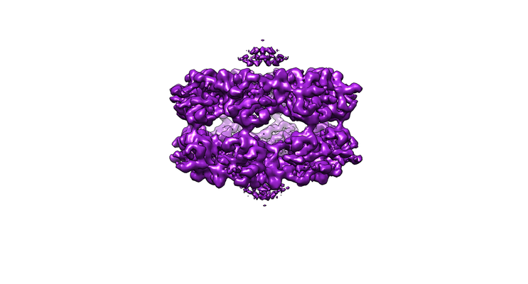 emd_3595.png
 http://ftp.pdbj.org/pub/emdb/structures/EMD-3595


 Z (Sec.)
 Y (Row.)
 X (Col.)


 Sample components
 Processing
 Electron microscopy
 FIELD EMISSION GUN

