+ Open data
- Basic information
| Entry | Database: EMDB / ID: EMD-3385 | |||||||||
|---|---|---|---|---|---|---|---|---|---|---|
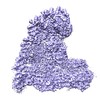
| Title | Cryo-EM structure of the human APC/C-Cdc20-Hsl1 complex | |||||||||
 Map data Map data | Single particle reconstruction of the human APC/C-Cdc20-Hsl1 complex | |||||||||
 Sample Sample |
| |||||||||
 Keywords Keywords | Cell cycle / phosphorylation / mitosis / ubiquitination | |||||||||
| Function / homology |  Function and homology information Function and homology informationnegative regulation of diacylglycerol biosynthetic process / protein localization to septin ring / mitotic morphogenesis checkpoint signaling / cellular bud neck septin ring / metaphase/anaphase transition of cell cycle / metaphase/anaphase transition of meiosis I / Inhibition of the proteolytic activity of APC/C required for the onset of anaphase by mitotic spindle checkpoint components / mitotic checkpoint complex / positive regulation of anaphase-promoting complex-dependent catabolic process / positive regulation of synapse maturation ...negative regulation of diacylglycerol biosynthetic process / protein localization to septin ring / mitotic morphogenesis checkpoint signaling / cellular bud neck septin ring / metaphase/anaphase transition of cell cycle / metaphase/anaphase transition of meiosis I / Inhibition of the proteolytic activity of APC/C required for the onset of anaphase by mitotic spindle checkpoint components / mitotic checkpoint complex / positive regulation of anaphase-promoting complex-dependent catabolic process / positive regulation of synapse maturation / regulation of meiotic nuclear division / Conversion from APC/C:Cdc20 to APC/C:Cdh1 in late anaphase / regulation of mitotic cell cycle spindle assembly checkpoint / Inactivation of APC/C via direct inhibition of the APC/C complex / APC/C:Cdc20 mediated degradation of mitotic proteins / regulation of dendrite development / positive regulation of synaptic plasticity / anaphase-promoting complex / Phosphorylation of Emi1 / regulation of meiotic cell cycle / Aberrant regulation of mitotic exit in cancer due to RB1 defects / anaphase-promoting complex-dependent catabolic process / protein branched polyubiquitination / metaphase/anaphase transition of mitotic cell cycle / Phosphorylation of the APC/C / anaphase-promoting complex binding / regulation of exit from mitosis / positive regulation of mitotic metaphase/anaphase transition / cellular bud neck / positive regulation of ubiquitin protein ligase activity / ubiquitin ligase activator activity / positive regulation of dendrite morphogenesis / protein K11-linked ubiquitination / regulation of mitotic metaphase/anaphase transition / mitotic sister chromatid cohesion / ubiquitin-ubiquitin ligase activity / mitotic metaphase chromosome alignment / mitotic spindle assembly checkpoint signaling / Regulation of APC/C activators between G1/S and early anaphase / Transcriptional Regulation by VENTX / cullin family protein binding / mitotic spindle assembly / enzyme-substrate adaptor activity / positive regulation of axon extension / protein K48-linked ubiquitination / ubiquitin-like ligase-substrate adaptor activity / intercellular bridge / heterochromatin / Amplification of signal from unattached kinetochores via a MAD2 inhibitory signal / nuclear periphery / Mitotic Prometaphase / APC/C:Cdc20 mediated degradation of Cyclin B / EML4 and NUDC in mitotic spindle formation / regulation of mitotic cell cycle / APC-Cdc20 mediated degradation of Nek2A / Resolution of Sister Chromatid Cohesion / Autodegradation of Cdh1 by Cdh1:APC/C / APC/C:Cdc20 mediated degradation of Securin / protein catabolic process / SCF-beta-TrCP mediated degradation of Emi1 / Assembly of the pre-replicative complex / Cdc20:Phospho-APC/C mediated degradation of Cyclin A / RHO GTPases Activate Formins / APC/C:Cdh1 mediated degradation of Cdc20 and other APC/C:Cdh1 targeted proteins in late mitosis/early G1 / G protein-coupled receptor binding / brain development / kinetochore / CDK-mediated phosphorylation and removal of Cdc6 / G2/M transition of mitotic cell cycle / spindle / histone deacetylase binding / neuron projection development / spindle pole / ubiquitin-protein transferase activity / mitotic spindle / Separation of Sister Chromatids / ubiquitin protein ligase activity / nervous system development / mitotic cell cycle / Antigen processing: Ubiquitination & Proteasome degradation / microtubule cytoskeleton / Senescence-Associated Secretory Phenotype (SASP) / protein phosphatase binding / ubiquitin-dependent protein catabolic process / molecular adaptor activity / cell differentiation / protein kinase activity / non-specific serine/threonine protein kinase / regulation of cell cycle / Ub-specific processing proteases / protein ubiquitination / negative regulation of gene expression / cell division / protein serine kinase activity / protein serine/threonine kinase activity / positive regulation of cell population proliferation / ubiquitin protein ligase binding / centrosome / nucleolus / perinuclear region of cytoplasm Similarity search - Function | |||||||||
| Biological species |  Homo sapiens (human) / Homo sapiens (human) /  | |||||||||
| Method | single particle reconstruction / cryo EM / negative staining / Resolution: 3.9 Å | |||||||||
 Authors Authors | Zhang S / Chang L / Alfieri C / Zhang Z / Yang J / Maslen S / Skehel M / Barford D | |||||||||
 Citation Citation |  Journal: Nature / Year: 2016 Journal: Nature / Year: 2016Title: Molecular mechanism of APC/C activation by mitotic phosphorylation. Authors: Suyang Zhang / Leifu Chang / Claudio Alfieri / Ziguo Zhang / Jing Yang / Sarah Maslen / Mark Skehel / David Barford /  Abstract: In eukaryotes, the anaphase-promoting complex (APC/C, also known as the cyclosome) regulates the ubiquitin-dependent proteolysis of specific cell-cycle proteins to coordinate chromosome segregation ...In eukaryotes, the anaphase-promoting complex (APC/C, also known as the cyclosome) regulates the ubiquitin-dependent proteolysis of specific cell-cycle proteins to coordinate chromosome segregation in mitosis and entry into the G1 phase. The catalytic activity of the APC/C and its ability to specify the destruction of particular proteins at different phases of the cell cycle are controlled by its interaction with two structurally related coactivator subunits, Cdc20 and Cdh1. Coactivators recognize substrate degrons, and enhance the affinity of the APC/C for its cognate E2 (refs 4-6). During mitosis, cyclin-dependent kinase (Cdk) and polo-like kinase (Plk) control Cdc20- and Cdh1-mediated activation of the APC/C. Hyperphosphorylation of APC/C subunits, notably Apc1 and Apc3, is required for Cdc20 to activate the APC/C, whereas phosphorylation of Cdh1 prevents its association with the APC/C. Since both coactivators associate with the APC/C through their common C-box and Ile-Arg tail motifs, the mechanism underlying this differential regulation is unclear, as is the role of specific APC/C phosphorylation sites. Here, using cryo-electron microscopy and biochemical analysis, we define the molecular basis of how phosphorylation of human APC/C allows for its control by Cdc20. An auto-inhibitory segment of Apc1 acts as a molecular switch that in apo unphosphorylated APC/C interacts with the C-box binding site and obstructs engagement of Cdc20. Phosphorylation of the auto-inhibitory segment displaces it from the C-box-binding site. Efficient phosphorylation of the auto-inhibitory segment, and thus relief of auto-inhibition, requires the recruitment of Cdk-cyclin in complex with a Cdk regulatory subunit (Cks) to a hyperphosphorylated loop of Apc3. We also find that the small-molecule inhibitor, tosyl-l-arginine methyl ester, preferentially suppresses APC/C(Cdc20) rather than APC/C(Cdh1), and interacts with the binding sites of both the C-box and Ile-Arg tail motifs. Our results reveal the mechanism for the regulation of mitotic APC/C by phosphorylation and provide a rationale for the development of selective inhibitors of this state. | |||||||||
| History |
|
- Structure visualization
| Movie |
 Movie viewer Movie viewer |
|---|---|
| Structure viewer | EM map:  SurfView SurfView Molmil Molmil Jmol/JSmol Jmol/JSmol |
| Supplemental images |
- Downloads & links
-EMDB archive
| Map data |  emd_3385.map.gz emd_3385.map.gz | 10 MB |  EMDB map data format EMDB map data format | |
|---|---|---|---|---|
| Header (meta data) |  emd-3385-v30.xml emd-3385-v30.xml emd-3385.xml emd-3385.xml | 26.1 KB 26.1 KB | Display Display |  EMDB header EMDB header |
| Images |  EMD-3385.jpg EMD-3385.jpg | 76.9 KB | ||
| Archive directory |  http://ftp.pdbj.org/pub/emdb/structures/EMD-3385 http://ftp.pdbj.org/pub/emdb/structures/EMD-3385 ftp://ftp.pdbj.org/pub/emdb/structures/EMD-3385 ftp://ftp.pdbj.org/pub/emdb/structures/EMD-3385 | HTTPS FTP |
-Related structure data
| Related structure data |  5g04MC  3386C  3387C  3388C  3389C  3390C  5g05C M: atomic model generated by this map C: citing same article ( |
|---|---|
| Similar structure data |
- Links
Links
| EMDB pages |  EMDB (EBI/PDBe) / EMDB (EBI/PDBe) /  EMDataResource EMDataResource |
|---|---|
| Related items in Molecule of the Month |
- Map
Map
| File |  Download / File: emd_3385.map.gz / Format: CCP4 / Size: 85.3 MB / Type: IMAGE STORED AS FLOATING POINT NUMBER (4 BYTES) Download / File: emd_3385.map.gz / Format: CCP4 / Size: 85.3 MB / Type: IMAGE STORED AS FLOATING POINT NUMBER (4 BYTES) | ||||||||||||||||||||||||||||||||||||||||||||||||||||||||||||
|---|---|---|---|---|---|---|---|---|---|---|---|---|---|---|---|---|---|---|---|---|---|---|---|---|---|---|---|---|---|---|---|---|---|---|---|---|---|---|---|---|---|---|---|---|---|---|---|---|---|---|---|---|---|---|---|---|---|---|---|---|---|
| Annotation | Single particle reconstruction of the human APC/C-Cdc20-Hsl1 complex | ||||||||||||||||||||||||||||||||||||||||||||||||||||||||||||
| Projections & slices | Image control
Images are generated by Spider. | ||||||||||||||||||||||||||||||||||||||||||||||||||||||||||||
| Voxel size | X=Y=Z: 1.36 Å | ||||||||||||||||||||||||||||||||||||||||||||||||||||||||||||
| Density |
| ||||||||||||||||||||||||||||||||||||||||||||||||||||||||||||
| Symmetry | Space group: 1 | ||||||||||||||||||||||||||||||||||||||||||||||||||||||||||||
| Details | EMDB XML:
CCP4 map header:
| ||||||||||||||||||||||||||||||||||||||||||||||||||||||||||||
-Supplemental data
- Sample components
+Entire : Recombinant human APC/C-Cdc20-Hsl1 complex
+Supramolecule #1000: Recombinant human APC/C-Cdc20-Hsl1 complex
+Macromolecule #1: Anaphase-promoting complex subunit 1
+Macromolecule #2: Anaphase-promoting complex subunit 2
+Macromolecule #3: Anaphase-promoting complex subunit 3
+Macromolecule #4: Anaphase-promoting complex subunit 4
+Macromolecule #5: Anaphase-promoting complex subunit 5
+Macromolecule #6: Anaphase-promoting complex subunit 6
+Macromolecule #7: Anaphase-promoting complex subunit 7
+Macromolecule #8: Anaphase-promoting complex subunit 8
+Macromolecule #9: Anaphase-promoting complex subunit 10
+Macromolecule #10: Anaphase-promoting complex subunit 11
+Macromolecule #11: Anaphase-promoting complex subunit 12
+Macromolecule #12: Anaphase-promoting complex subunit 13
+Macromolecule #13: Anaphase-promoting complex subunit 15
+Macromolecule #14: Anaphase-promoting complex subunit 16
+Macromolecule #15: Cell division cycle protein 20 homolog
+Macromolecule #16: Hsl1
-Experimental details
-Structure determination
| Method | negative staining, cryo EM |
|---|---|
 Processing Processing | single particle reconstruction |
| Aggregation state | particle |
- Sample preparation
| Concentration | 0.15 mg/mL |
|---|---|
| Buffer | pH: 8 / Details: 20mM Hepes, 150mM NaCl, 0.005mM TCEP |
| Staining | Type: NEGATIVE / Details: Vitrification in liquid ethane |
| Grid | Details: 200 mesh Quantifoil R2/2 copper grid with thin carbon support, treated with a 9:1 argon:oxygen plasma cleaner for 20-40s |
| Vitrification | Cryogen name: ETHANE / Chamber humidity: 100 % / Chamber temperature: 100 K / Instrument: FEI VITROBOT MARK III Method: The grids were incubated for 30s at 4 degree and 100% humidity before blotting for 5s and plunging into liquid ethane |
- Electron microscopy
| Microscope | FEI POLARA 300 |
|---|---|
| Temperature | Min: 90 K / Max: 120 K / Average: 105 K |
| Specialist optics | Energy filter - Name: FEI |
| Date | Oct 30, 2015 |
| Image recording | Category: CCD / Film or detector model: FEI FALCON II (4k x 4k) / Number real images: 11000 / Average electron dose: 27 e/Å2 Details: The exposure time for each micrograph was 2s at a dose rate of 27 electrons/angstrom2/s. 34 movie frames were recorded for each micrograph. |
| Electron beam | Acceleration voltage: 300 kV / Electron source:  FIELD EMISSION GUN FIELD EMISSION GUN |
| Electron optics | Illumination mode: FLOOD BEAM / Imaging mode: BRIGHT FIELD / Cs: 2 mm / Nominal defocus max: 4.0 µm / Nominal defocus min: 2.0 µm / Nominal magnification: 78000 |
| Sample stage | Specimen holder: liquid nitrogen cooled / Specimen holder model: OTHER |
| Experimental equipment |  Model: Tecnai Polara / Image courtesy: FEI Company |
- Image processing
| Details | Particles were selected by automatic particle picking in RELION |
|---|---|
| CTF correction | Details: Each micrograph |
| Final reconstruction | Applied symmetry - Point group: C1 (asymmetric) / Resolution.type: BY AUTHOR / Resolution: 3.9 Å / Resolution method: OTHER / Software - Name: RELION / Number images used: 179660 |
 Movie
Movie Controller


 Z (Sec.)
 Y (Row.)
 X (Col.)


 baculovirus-insect cells
