+ Open data
- Basic information
| Entry | Database: EMDB / ID: EMD-3168 | |||||||||
|---|---|---|---|---|---|---|---|---|---|---|
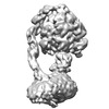
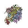
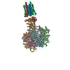
| Title | Bovine mitochondrial ATP synthase state 2c | |||||||||
 Map data Map data | Reconstruction of detergent-solubilized bovine mitochondrial ATP synthase | |||||||||
 Sample Sample |
| |||||||||
 Keywords Keywords | ATP synthase / rotary ATPase | |||||||||
| Function / homology |  Function and homology information Function and homology informationMitochondrial protein import / Formation of ATP by chemiosmotic coupling / Cristae formation / mitochondrial proton-transporting ATP synthase complex assembly / mitochondrial envelope / proton channel activity / Mitochondrial protein degradation / proton transmembrane transporter activity / proton motive force-driven ATP synthesis / proton-transporting two-sector ATPase complex, proton-transporting domain ...Mitochondrial protein import / Formation of ATP by chemiosmotic coupling / Cristae formation / mitochondrial proton-transporting ATP synthase complex assembly / mitochondrial envelope / proton channel activity / Mitochondrial protein degradation / proton transmembrane transporter activity / proton motive force-driven ATP synthesis / proton-transporting two-sector ATPase complex, proton-transporting domain / proton motive force-driven mitochondrial ATP synthesis / H+-transporting two-sector ATPase / proton-transporting ATP synthase complex / proton-transporting ATP synthase activity, rotational mechanism / proton transmembrane transport / aerobic respiration / ADP binding / mitochondrial membrane / mitochondrial inner membrane / lipid binding / structural molecule activity / ATP hydrolysis activity / mitochondrion / ATP binding / plasma membrane Similarity search - Function | |||||||||
| Biological species |  | |||||||||
| Method | single particle reconstruction / cryo EM / Resolution: 7.4 Å | |||||||||
 Authors Authors | Zhou A / Rohou A / Schep DG / Bason JV / Montgomery MG / Walker JE / Grigorieff N / Rubinstein JL | |||||||||
 Citation Citation |  Journal: Elife / Year: 2015 Journal: Elife / Year: 2015Title: Structure and conformational states of the bovine mitochondrial ATP synthase by cryo-EM. Authors: Anna Zhou / Alexis Rohou / Daniel G Schep / John V Bason / Martin G Montgomery / John E Walker / Nikolaus Grigorieff / John L Rubinstein /    Abstract: Adenosine triphosphate (ATP), the chemical energy currency of biology, is synthesized in eukaryotic cells primarily by the mitochondrial ATP synthase. ATP synthases operate by a rotary catalytic ...Adenosine triphosphate (ATP), the chemical energy currency of biology, is synthesized in eukaryotic cells primarily by the mitochondrial ATP synthase. ATP synthases operate by a rotary catalytic mechanism where proton translocation through the membrane-inserted FO region is coupled to ATP synthesis in the catalytic F1 region via rotation of a central rotor subcomplex. We report here single particle electron cryomicroscopy (cryo-EM) analysis of the bovine mitochondrial ATP synthase. Combining cryo-EM data with bioinformatic analysis allowed us to determine the fold of the a subunit, suggesting a proton translocation path through the FO region that involves both the a and b subunits. 3D classification of images revealed seven distinct states of the enzyme that show different modes of bending and twisting in the intact ATP synthase. Rotational fluctuations of the c8-ring within the FO region support a Brownian ratchet mechanism for proton-translocation-driven rotation in ATP synthases. | |||||||||
| History |
|
- Structure visualization
| Movie |
 Movie viewer Movie viewer |
|---|---|
| Structure viewer | EM map:  SurfView SurfView Molmil Molmil Jmol/JSmol Jmol/JSmol |
| Supplemental images |
- Downloads & links
-EMDB archive
| Map data |  emd_3168.map.gz emd_3168.map.gz | 51.1 MB |  EMDB map data format EMDB map data format | |
|---|---|---|---|---|
| Header (meta data) |  emd-3168-v30.xml emd-3168-v30.xml emd-3168.xml emd-3168.xml | 16.2 KB 16.2 KB | Display Display |  EMDB header EMDB header |
| Images |  EMD-3168.tif EMD-3168.tif | 129.2 KB | ||
| Archive directory |  http://ftp.pdbj.org/pub/emdb/structures/EMD-3168 http://ftp.pdbj.org/pub/emdb/structures/EMD-3168 ftp://ftp.pdbj.org/pub/emdb/structures/EMD-3168 ftp://ftp.pdbj.org/pub/emdb/structures/EMD-3168 | HTTPS FTP |
-Related structure data
| Related structure data |  5fijMC  3164C  3165C  3166C  3167C  3169C  3170C  3181C  5araC  5areC  5arhC  5ariC  5fikC  5filC M: atomic model generated by this map C: citing same article ( |
|---|---|
| Similar structure data |
- Links
Links
| EMDB pages |  EMDB (EBI/PDBe) / EMDB (EBI/PDBe) /  EMDataResource EMDataResource |
|---|---|
| Related items in Molecule of the Month |
- Map
Map
| File |  Download / File: emd_3168.map.gz / Format: CCP4 / Size: 62.5 MB / Type: IMAGE STORED AS FLOATING POINT NUMBER (4 BYTES) Download / File: emd_3168.map.gz / Format: CCP4 / Size: 62.5 MB / Type: IMAGE STORED AS FLOATING POINT NUMBER (4 BYTES) | ||||||||||||||||||||||||||||||||||||||||||||||||||||||||||||
|---|---|---|---|---|---|---|---|---|---|---|---|---|---|---|---|---|---|---|---|---|---|---|---|---|---|---|---|---|---|---|---|---|---|---|---|---|---|---|---|---|---|---|---|---|---|---|---|---|---|---|---|---|---|---|---|---|---|---|---|---|---|
| Annotation | Reconstruction of detergent-solubilized bovine mitochondrial ATP synthase | ||||||||||||||||||||||||||||||||||||||||||||||||||||||||||||
| Projections & slices | Image control
Images are generated by Spider. | ||||||||||||||||||||||||||||||||||||||||||||||||||||||||||||
| Voxel size | X=Y=Z: 1.64 Å | ||||||||||||||||||||||||||||||||||||||||||||||||||||||||||||
| Density |
| ||||||||||||||||||||||||||||||||||||||||||||||||||||||||||||
| Symmetry | Space group: 1 | ||||||||||||||||||||||||||||||||||||||||||||||||||||||||||||
| Details | EMDB XML:
CCP4 map header:
| ||||||||||||||||||||||||||||||||||||||||||||||||||||||||||||
-Supplemental data
- Sample components
-Entire : Bovine mitochondrial ATP synthase
| Entire | Name: Bovine mitochondrial ATP synthase |
|---|---|
| Components |
|
-Supramolecule #1000: Bovine mitochondrial ATP synthase
| Supramolecule | Name: Bovine mitochondrial ATP synthase / type: sample / ID: 1000 / Details: Detergent-solubilized protein complex Oligomeric state: One hetero-oligomeric ATP synthase complex Number unique components: 1 |
|---|---|
| Molecular weight | Experimental: 600 KDa |
-Macromolecule #1: ATP synthase
| Macromolecule | Name: ATP synthase / type: protein_or_peptide / ID: 1 / Name.synonym: ATPase, complex V / Number of copies: 1 / Oligomeric state: monomer / Recombinant expression: No |
|---|---|
| Source (natural) | Organism:  |
| Molecular weight | Experimental: 600 KDa |
-Experimental details
-Structure determination
| Method | cryo EM |
|---|---|
 Processing Processing | single particle reconstruction |
| Aggregation state | particle |
- Sample preparation
| Concentration | 8 mg/mL |
|---|---|
| Buffer | pH: 7.2 Details: 20 mM Tris-HCl, 100 mM NaCl, 0.05% (wt/v) dodecylmaltoside, 2 mM ATP, 0.02% (wt/v) NaN3 |
| Grid | Details: Homemade holey carbon on 400 square mesh Cu/Rh grid, glow-discharged 2 mins |
| Vitrification | Cryogen name: ETHANE-PROPANE MIXTURE / Chamber humidity: 100 % / Instrument: FEI VITROBOT MARK III / Method: Blot for 27 seconds before plunging |
- Electron microscopy #1
| Microscopy ID | 1 |
|---|---|
| Microscope | FEI TITAN KRIOS |
| Temperature | Average: 80 K |
| Alignment procedure | Legacy - Astigmatism: Objective lens astigmatism was corrected at 18,000x magnification (before detector post-magnification) |
| Details | K2 Summit direct detector device (Gatan Inc.) operated in super-resolution mode with a 1.64 angstrom physical pixel and 0.82 angstrom super-resolution pixel. With no specimen present, the rate of exposure of the detector was 8 electrons/pixel/second. Exposure- fractionated movies of 20.1 s were recorded as stacks of 67 frames, so that selected specimen areas were exposed with a total of 60.3 electrons/square angstrom. |
| Date | Mar 15, 2015 |
| Image recording | Category: CCD / Film or detector model: GATAN K2 (4k x 4k) / Digitization - Sampling interval: 5 µm / Number real images: 5867 / Average electron dose: 60.3 e/Å2 / Bits/pixel: 8 |
| Electron beam | Acceleration voltage: 300 kV / Electron source:  FIELD EMISSION GUN FIELD EMISSION GUN |
| Electron optics | Calibrated magnification: 30487 / Illumination mode: FLOOD BEAM / Imaging mode: BRIGHT FIELD / Cs: 2.7 mm / Nominal defocus max: 4.1 µm / Nominal defocus min: 1.2 µm / Nominal magnification: 18000 |
| Sample stage | Specimen holder model: FEI TITAN KRIOS AUTOGRID HOLDER |
| Experimental equipment |  Model: Titan Krios / Image courtesy: FEI Company |
- Electron microscopy #2
| Microscopy ID | 2 |
|---|---|
| Microscope | FEI TITAN KRIOS |
| Temperature | Average: 80 K |
| Alignment procedure | Legacy - Astigmatism: Objective lens astigmatism was corrected at 18,000x magnification (before detector post-magnification) |
| Details | K2 Summit direct detector device (Gatan Inc.) operated in super-resolution mode with a 1.64 angstrom physical pixel and 0.82 angstrom super-resolution pixel. With no specimen present, the rate of exposure of the detector was 8 electrons/pixel/second. Exposure- fractionated movies of 20.1 s were recorded as stacks of 67 frames, so that selected specimen areas were exposed with a total of 60.3 electrons/square angstrom. |
| Date | Sep 28, 2014 |
| Image recording | Category: CCD / Film or detector model: GATAN K2 (4k x 4k) / Digitization - Sampling interval: 5 µm / Number real images: 5867 / Average electron dose: 60.3 e/Å2 / Bits/pixel: 8 |
| Electron beam | Acceleration voltage: 300 kV / Electron source:  FIELD EMISSION GUN FIELD EMISSION GUN |
| Electron optics | Calibrated magnification: 30487 / Illumination mode: FLOOD BEAM / Imaging mode: BRIGHT FIELD / Cs: 2.7 mm / Nominal defocus max: 4.1 µm / Nominal defocus min: 1.2 µm / Nominal magnification: 18000 |
| Sample stage | Specimen holder model: FEI TITAN KRIOS AUTOGRID HOLDER |
| Experimental equipment |  Model: Titan Krios / Image courtesy: FEI Company |
- Image processing
| Details | The particles were selected using an automatic selection program. |
|---|---|
| CTF correction | Details: Each particle |
| Final reconstruction | Applied symmetry - Point group: C1 (asymmetric) / Algorithm: OTHER / Resolution.type: BY AUTHOR / Resolution: 7.4 Å / Resolution method: OTHER / Software - Name: Relion, FREALIGN Details: To avoid noise bias, only data up to a resolution of 10 angstrom were used during refinement. Number images used: 18899 |
-Atomic model buiding 1
| Initial model | PDB ID: Chain - #0 - Chain ID: A / Chain - #1 - Chain ID: B / Chain - #2 - Chain ID: C / Chain - #3 - Chain ID: D / Chain - #4 - Chain ID: E / Chain - #5 - Chain ID: F / Chain - #6 - Chain ID: G / Chain - #7 - Chain ID: H / Chain - #8 - Chain ID: I / Chain - #9 - Chain ID: S |
|---|---|
| Software | Name: Chimera, MDFF |
| Details | Rigid body fitting performed in Chimera first, followed by flexible fitting performed using Molecular Dynamics Flexible Fitting (MDFF). |
| Refinement | Space: REAL / Protocol: FLEXIBLE FIT |
| Output model |  PDB-5fij: |
-Atomic model buiding 2
| Initial model | PDB ID: Chain - #0 - Chain ID: J / Chain - #1 - Chain ID: K / Chain - #2 - Chain ID: L / Chain - #3 - Chain ID: M / Chain - #4 - Chain ID: N / Chain - #5 - Chain ID: O / Chain - #6 - Chain ID: P / Chain - #7 - Chain ID: Q |
|---|---|
| Software | Name: Chimera, MDFF |
| Details | Rigid body fitting performed in Chimera first, followed by flexible fitting performed using Molecular Dynamics Flexible Fitting (MDFF). |
| Refinement | Space: REAL / Protocol: FLEXIBLE FIT |
| Output model |  PDB-5fij: |
-Atomic model buiding 3
| Initial model | PDB ID: Chain - #0 - Chain ID: D / Chain - #1 - Chain ID: E / Chain - #2 - Chain ID: F |
|---|---|
| Software | Name: Chimera, MDFF |
| Details | Rigid body fitting performed in Chimera first, followed by flexible fitting performed using Molecular Dynamics Flexible Fitting (MDFF). |
| Refinement | Space: REAL / Protocol: FLEXIBLE FIT |
| Output model |  PDB-5fij: |
 Movie
Movie Controller


 Z (Sec.)
 Y (Row.)
 X (Col.)


