Movie
Movie Controller


+ Open data

- Basic information
| Entry | Database: PDB / ID: 5are | ||||||
|---|---|---|---|---|---|---|---|
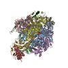
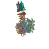
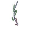
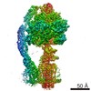
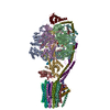
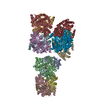
| Title | Bovine mitochondrial ATP synthase state 1b | ||||||
 Components Components |
| ||||||
 Keywords Keywords | HYDROLASE / ATP SYNTHASE / ROTARY ATPASE | ||||||
| Function / homology |  Function and homology information Function and homology informationMitochondrial protein import / Formation of ATP by chemiosmotic coupling / Cristae formation / mitochondrial proton-transporting ATP synthase complex assembly / mitochondrial envelope / Mitochondrial translation termination / proton channel activity / Mitochondrial protein degradation / proton transmembrane transporter activity / proton motive force-driven ATP synthesis ...Mitochondrial protein import / Formation of ATP by chemiosmotic coupling / Cristae formation / mitochondrial proton-transporting ATP synthase complex assembly / mitochondrial envelope / Mitochondrial translation termination / proton channel activity / Mitochondrial protein degradation / proton transmembrane transporter activity / proton motive force-driven ATP synthesis / proton-transporting two-sector ATPase complex, proton-transporting domain / proton motive force-driven mitochondrial ATP synthesis / H+-transporting two-sector ATPase / proton-transporting ATP synthase complex / proton-transporting ATP synthase activity, rotational mechanism / proton transmembrane transport / aerobic respiration / ADP binding / mitochondrial membrane / mitochondrial inner membrane / lipid binding / structural molecule activity / ATP hydrolysis activity / mitochondrion / ATP binding / metal ion binding / plasma membrane Similarity search - Function | ||||||
| Biological species |  | ||||||
| Method | ELECTRON MICROSCOPY / single particle reconstruction / cryo EM / Resolution: 6.7 Å | ||||||
 Authors Authors | Zhou, A. / Rohou, A. / Schep, D.G. / Bason, J.V. / Montgomery, M.G. / Walker, J.E. / Grigorieff, N. / Rubinstein, J.L. | ||||||
 Citation Citation | Journal: Elife / Year: 2015 Title: Structure and conformational states of the bovine mitochondrial ATP synthase by cryo-EM. Authors: Anna Zhou / Alexis Rohou / Daniel G Schep / John V Bason / Martin G Montgomery / John E Walker / Nikolaus Grigorieff / John L Rubinstein /    Abstract: Adenosine triphosphate (ATP), the chemical energy currency of biology, is synthesized in eukaryotic cells primarily by the mitochondrial ATP synthase. ATP synthases operate by a rotary catalytic ...Adenosine triphosphate (ATP), the chemical energy currency of biology, is synthesized in eukaryotic cells primarily by the mitochondrial ATP synthase. ATP synthases operate by a rotary catalytic mechanism where proton translocation through the membrane-inserted FO region is coupled to ATP synthesis in the catalytic F1 region via rotation of a central rotor subcomplex. We report here single particle electron cryomicroscopy (cryo-EM) analysis of the bovine mitochondrial ATP synthase. Combining cryo-EM data with bioinformatic analysis allowed us to determine the fold of the a subunit, suggesting a proton translocation path through the FO region that involves both the a and b subunits. 3D classification of images revealed seven distinct states of the enzyme that show different modes of bending and twisting in the intact ATP synthase. Rotational fluctuations of the c8-ring within the FO region support a Brownian ratchet mechanism for proton-translocation-driven rotation in ATP synthases. | ||||||
| History |
| ||||||
| Remark 700 | SHEET DETERMINATION METHOD: DSSP THE SHEETS PRESENTED AS "AA" IN EACH CHAIN ON SHEET RECORDS BELOW ... SHEET DETERMINATION METHOD: DSSP THE SHEETS PRESENTED AS "AA" IN EACH CHAIN ON SHEET RECORDS BELOW IS ACTUALLY AN 6-STRANDED BARREL THIS IS REPRESENTED BY A 7-STRANDED SHEET IN WHICH THE FIRST AND LAST STRANDS ARE IDENTICAL. SHEET DETERMINATION METHOD: DSSP THE SHEETS PRESENTED AS "BA" IN EACH CHAIN ON SHEET RECORDS BELOW IS ACTUALLY AN **-STRANDED BARREL THIS IS REPRESENTED BY A **-STRANDED SHEET IN WHICH THE FIRST AND LAST STRANDS ARE IDENTICAL. SHEET DETERMINATION METHOD: DSSP THE SHEETS PRESENTED AS "DA" IN EACH CHAIN ON SHEET RECORDS BELOW IS ACTUALLY AN 0-STRANDED BARREL THIS IS REPRESENTED BY A 1-STRANDED SHEET IN WHICH THE FIRST AND LAST STRANDS ARE IDENTICAL. SHEET DETERMINATION METHOD: DSSP THE SHEETS PRESENTED AS "EA" IN EACH CHAIN ON SHEET RECORDS BELOW IS ACTUALLY AN 0-STRANDED BARREL THIS IS REPRESENTED BY A 1-STRANDED SHEET IN WHICH THE FIRST AND LAST STRANDS ARE IDENTICAL. SHEET THE SHEET STRUCTURE OF THIS MOLECULE IS BIFURCATED. IN ORDER TO REPRESENT THIS FEATURE IN THE SHEET RECORDS BELOW, TWO SHEETS ARE DEFINED. |
- Structure visualization
| Movie |
Movie viewer |
|---|---|
| Structure viewer | Molecule: MolmilJmol/JSmol |
- Downloads & links
-Download
| PDBx/mmCIF format | 5are.cif.gz | 565.1 KB | Display | PDBx/mmCIF format |
|---|---|---|---|---|
| PDB format | pdb5are.ent.gz | 349.4 KB | Display | PDB format |
| PDBx/mmJSON format | 5are.json.gz | Tree view | PDBx/mmJSON format | |
| Others |  Other downloads Other downloads |
-Validation report
| Arichive directory | https://data.pdbj.org/pub/pdb/validation_reports/ar/5areftp://data.pdbj.org/pub/pdb/validation_reports/ar/5are | HTTPS FTP |
|---|
-Related structure data
| Related structure data | 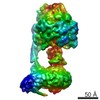 3165MC 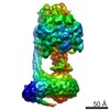 3164C 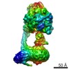 3166C 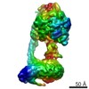 3167C 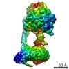 3168C 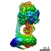 3169C 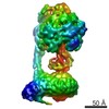 3170C 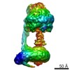 3181C 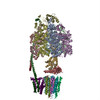 5araC 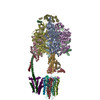 5arhC 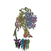 5ariC  5fijC  5fikC 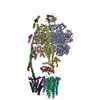 5filC C: citing same article ( M: map data used to model this data |
|---|---|
| Similar structure data |


-Links
 PDBj
PDBj


- Assembly
| Deposited unit | 
|
|---|---|
| 1 |
|
-Components
-ATP SYNTHASE SUBUNIT ... , 8 types, 12 molecules ABCDEFGHISUW
| #1: Protein | Mass: 55301.207 Da / Num. of mol.: 3 / Fragment: UNP RESIDUES 44-553 / Source method: isolated from a natural source / Source: (natural) #2: Protein | Mass: 51757.836 Da / Num. of mol.: 3 / Fragment: UNP RESIDUES 47-528 / Source method: isolated from a natural source / Source: (natural) References: UniProt: P00829, H+-transporting two-sector ATPase #3: Protein | | Mass: 30300.760 Da / Num. of mol.: 1 / Fragment: UNP RESIDUES 26-298 / Source method: isolated from a natural source / Source: (natural) #4: Protein | | Mass: 15074.813 Da / Num. of mol.: 1 / Fragment: UNP RESIDUES 23-168 / Source method: isolated from a natural source / Source: (natural) #5: Protein/peptide | | Mass: 5662.693 Da / Num. of mol.: 1 / Fragment: UNP RESIDUES 2-51 / Source method: isolated from a natural source / Source: (natural) #7: Protein | | Mass: 20989.803 Da / Num. of mol.: 1 / Fragment: UNP RESIDUES 24-213 Source method: isolated from a genetically manipulated source Source: (gene. exp.)  #9: Protein | | Mass: 14167.169 Da / Num. of mol.: 1 / Fragment: UNP RESIDUES 2-125 Source method: isolated from a genetically manipulated source Source: (gene. exp.) #11: Protein | | Mass: 23717.578 Da / Num. of mol.: 1 / Fragment: UNP RESIDUES 10-226 / Source method: isolated from a natural source / Source: (natural) |
|---|
-ATP SYNTHASE F(0) COMPLEX SUBUNIT ... , 2 types, 9 molecules JKLMNOPQT
| #6: Protein | Mass: 7293.593 Da / Num. of mol.: 8 / Fragment: UNP RESIDUES 63-134 / Source method: isolated from a natural source / Source: (natural) #8: Protein | | Mass: 20335.625 Da / Num. of mol.: 1 / Fragment: UNP RESIDUES 76-249 Source method: isolated from a genetically manipulated source Source: (gene. exp.) |
|---|
-Protein , 1 types, 1 molecules V
| #10: Protein | Mass: 9118.253 Da / Num. of mol.: 1 / Fragment: UNP RESIDUES 32-108 Source method: isolated from a genetically manipulated source Source: (gene. exp.) |
|---|
-Experimental details
-Experiment
| Experiment | Method: ELECTRON MICROSCOPY |
|---|---|
| EM experiment | Aggregation state: PARTICLE / 3D reconstruction method: single particle reconstruction |
- Sample preparation
| Component | Name: BOVINE MITOCHONDRIAL ATP SYNTHASE / Type: COMPLEX |
|---|---|
| Buffer solution | Name: 20 MM TRIS-HCL, 100 MM NACL, 10% (V/V) DODECYLMALTOSIDE, 2 MM ATP, 0.02% (WT/V) NAN3 pH: 7.2 Details: 20 MM TRIS-HCL, 100 MM NACL, 10% (V/V) DODECYLMALTOSIDE, 2 MM ATP, 0.02% (WT/V) NAN3 |
| Specimen | Conc.: 8 mg/ml / Embedding applied: NO / Shadowing applied: NO / Staining applied: NO / Vitrification applied: YES |
| Specimen support | Details: HOLEY CARBON |
| Vitrification | Instrument: FEI VITROBOT MARK III / Cryogen name: ETHANE-PROPANE Details: VITRIFICATION 1 -- CRYOGEN- ETHANE-PROPANE MIXTURE, HUMIDITY- 100, INSTRUMENT- FEI VITROBOT MARK III, METHOD- BLOT FOR 27 SECONDS BEFORE PLUNGING, |
- Electron microscopy imaging
| Experimental equipment |  Model: Titan Krios / Image courtesy: FEI Company |
|---|---|
| Microscopy | Model: FEI TITAN KRIOS / Date: Mar 15, 2015 Details: K2 SUMMIT DIRECT DETECTOR DEVICE (GATAN INC.) OPERATED IN SUPER- RESOLUTION MODE WITH A 1.64 ANGSTROM PHYSICAL PIXEL AND 0. 82 ANGSTROM SUPER- RESOLUTION PIXEL. WITH NO SPECIMEN PRESENT, THE ...Details: K2 SUMMIT DIRECT DETECTOR DEVICE (GATAN INC.) OPERATED IN SUPER- RESOLUTION MODE WITH A 1.64 ANGSTROM PHYSICAL PIXEL AND 0. 82 ANGSTROM SUPER- RESOLUTION PIXEL. WITH NO SPECIMEN PRESENT, THE RATE OF EXPOSURE OF THE DETECTOR WAS 8 ELECTRONS PER PIXEL PER SECOND. EXPOSURE- FRACTIONATED MOVIES OF 20.1 S WERE RECORDED AS STACKS OF 67 FRAMES, SO THAT SELECTED SPECIMEN AREAS WERE EXPOSED WITH A TOTAL OF 60.3 ELECTRONS PER SQUARE ANGSTROM. |
| Electron gun | Electron source:  FIELD EMISSION GUN / Accelerating voltage: 300 kV / Illumination mode: FLOOD BEAM FIELD EMISSION GUN / Accelerating voltage: 300 kV / Illumination mode: FLOOD BEAM |
| Electron lens | Mode: BRIGHT FIELD / Nominal magnification: 18000 X / Calibrated magnification: 30487 X / Nominal defocus max: 4100 nm / Nominal defocus min: 1200 nm / Cs: 2.7 mm |
| Specimen holder | Temperature: 80 K |
| Image recording | Electron dose: 60.3 e/Å2 / Film or detector model: GATAN K2 (4k x 4k) |
- Processing
| EM software |
| ||||||||||||||||||||||||||||
|---|---|---|---|---|---|---|---|---|---|---|---|---|---|---|---|---|---|---|---|---|---|---|---|---|---|---|---|---|---|
| CTF correction | Details: EACH PARTICLE | ||||||||||||||||||||||||||||
| Symmetry | Point symmetry: C1 (asymmetric) | ||||||||||||||||||||||||||||
| 3D reconstruction | Method: PROJECTION MATCHING AND MAXIMUM LIKELIHOOD CLASSIFICATION Resolution: 6.7 Å / Num. of particles: 22935 Details: THE A SUBUNIT WAS MODELED USING EVOLUTIONARY COVARIANCE. THE N-TERMINAL TRANSMEMBRANE HELICES OF THE B SUBUNIT WERE MODELED BASED ON TRANSMEMBRANE HELIX PREDICTIONS. SUBMISSION BASED ON ...Details: THE A SUBUNIT WAS MODELED USING EVOLUTIONARY COVARIANCE. THE N-TERMINAL TRANSMEMBRANE HELICES OF THE B SUBUNIT WERE MODELED BASED ON TRANSMEMBRANE HELIX PREDICTIONS. SUBMISSION BASED ON EXPERIMENTAL DATA FROM EMDB EMD-3165. (DEPOSITION ID: 13793). Symmetry type: POINT | ||||||||||||||||||||||||||||
| Atomic model building | Protocol: FLEXIBLE FIT / Details: METHOD--FLEXIBLE FITTING | ||||||||||||||||||||||||||||
| Atomic model building |
| ||||||||||||||||||||||||||||
| Refinement | Highest resolution: 6.7 Å | ||||||||||||||||||||||||||||
| Refinement step | Cycle: LAST / Highest resolution: 7.4 Å
|