 Movie
Movie Controller

[English] 日本語
 Yorodumi
- PDB-2x31: Modelling of the complex between subunits BchI and BchD of magnes... -
+ Open data

- Basic information
| Entry | Database: PDB / ID: 2x31 | ||||||
|---|---|---|---|---|---|---|---|
| Title | Modelling of the complex between subunits BchI and BchD of magnesium chelatase based on single-particle cryo-EM reconstruction at 7.5 ang | ||||||
 Components Components |
| ||||||
 Keywords Keywords | LIGASE / BACTERIOCHLOROPHYLL BIOSYNTHESIS / PHOTOSYNTHESIS | ||||||
| Function / homology |  Function and homology information Function and homology informationbacteriochlorophyll biosynthetic process / magnesium chelatase / magnesium chelatase activity / photosynthesis / ATP hydrolysis activity / ATP binding Similarity search - Function | ||||||
| Biological species |  RHODOBACTER CAPSULATUS (bacteria) RHODOBACTER CAPSULATUS (bacteria) | ||||||
| Method | ELECTRON MICROSCOPY / single particle reconstruction / cryo EM / Resolution: 7.5 Å | ||||||
 Authors Authors | Lunqvist, J. / Elmlund, H. / Peterson Wulff, R. / Berglund, L. / Elmlund, D. / Emanuelsson, C. / Hebert, H. / Willows, R.D. / Hansson, M. / Lindahl, M. / Al-Karadaghi, S. | ||||||
 Citation Citation | Journal: Structure / Year: 2010 Title: ATP-induced conformational dynamics in the AAA+ motor unit of magnesium chelatase. Authors: Joakim Lundqvist / Hans Elmlund / Ragna Peterson Wulff / Lisa Berglund / Dominika Elmlund / Cecilia Emanuelsson / Hans Hebert / Robert D Willows / Mats Hansson / Martin Lindahl / Salam Al-Karadaghi /  Abstract: Mg-chelatase catalyzes the first committed step of the chlorophyll biosynthetic pathway, the ATP-dependent insertion of Mg(2+) into protoporphyrin IX (PPIX). Here we report the reconstruction using ...Mg-chelatase catalyzes the first committed step of the chlorophyll biosynthetic pathway, the ATP-dependent insertion of Mg(2+) into protoporphyrin IX (PPIX). Here we report the reconstruction using single-particle cryo-electron microscopy of the complex between subunits BchD and BchI of Rhodobacter capsulatus Mg-chelatase in the presence of ADP, the nonhydrolyzable ATP analog AMPPNP, and ATP at 7.5 A, 14 A, and 13 A resolution, respectively. We show that the two AAA+ modules of the subunits form a unique complex of 3 dimers related by a three-fold axis. The reconstructions demonstrate substantial differences between the conformations of the complex in the presence of ATP and ADP, and suggest that the C-terminal integrin-I domains of the BchD subunits play a central role in transmitting conformational changes of BchI to BchD. Based on these data a model for the function of magnesium chelatase is proposed. | ||||||
| History |
|
- Structure visualization
| Movie |
Movie viewer |
|---|---|
| Structure viewer | Molecule: MolmilJmol/JSmol |
- Downloads & links
-Download
| PDBx/mmCIF format | 2x31.cif.gz | 528.7 KB | Display | PDBx/mmCIF format |
|---|---|---|---|---|
| PDB format | pdb2x31.ent.gz | 430.2 KB | Display | PDB format |
| PDBx/mmJSON format | 2x31.json.gz | Tree view | PDBx/mmJSON format | |
| Others |  Other downloads Other downloads |
-Validation report
| Summary document | 2x31_validation.pdf.gz | 901.6 KB | Display | wwPDB validaton report |
|---|---|---|---|---|
| Full document | 2x31_full_validation.pdf.gz | 991.6 KB | Display | |
| Data in XML | 2x31_validation.xml.gz | 88.9 KB | Display | |
| Data in CIF | 2x31_validation.cif.gz | 130.5 KB | Display | |
| Arichive directory | https://data.pdbj.org/pub/pdb/validation_reports/x3/2x31ftp://data.pdbj.org/pub/pdb/validation_reports/x3/2x31 | HTTPS FTP |
-Related structure data
| Related structure data | 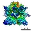 1676MC 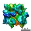 1677MC  1678MC M: map data used to model this data C: citing same article ( |
|---|---|
| Similar structure data |


-Links
 PDBj
PDBj


- Assembly
| Deposited unit | 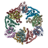
|
|---|---|
| 1 |
|
-Components
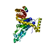
| #1: Protein | Mass: 19666.758 Da / Num. of mol.: 6 / Fragment: RESIDUES 373-561 Source method: isolated from a genetically manipulated source Source: (gene. exp.) RHODOBACTER CAPSULATUS (bacteria) / Production host: #2: Protein | Mass: 37946.340 Da / Num. of mol.: 6 Source method: isolated from a genetically manipulated source Source: (gene. exp.) RHODOBACTER CAPSULATUS (bacteria) / Production host: Sequence details | HOMOLOGY MODEL OF INTEGRIN I DOMAINS OF SUBUBUNIT BCHD A,B,C,D,E,F SUBUNITS BCHI G,H,I,J,K,L BASED ...HOMOLOGY MODEL OF INTEGRIN I DOMAINS OF SUBUBUNIT BCHD A,B,C,D,E,F SUBUNITS BCHI G,H,I,J,K,L BASED ON PDB ENTRY 1G8P | |
|---|
-Experimental details
-Experiment
| Experiment | Method: ELECTRON MICROSCOPY |
|---|---|
| EM experiment | Aggregation state: PARTICLE / 3D reconstruction method: single particle reconstruction |
- Sample preparation
| Component | Name: BCHID COMPLEX OF RHODOBACTER CAPSULATUS MG CHELATASE / Type: COMPLEX |
|---|---|
| Buffer solution | pH: 8 |
| Specimen | Conc.: 0.1 mg/ml / Embedding applied: NO / Shadowing applied: NO / Staining applied: NO / Vitrification applied: YES |
| Specimen support | Details: HOLEY CARBON |
| Vitrification | Cryogen name: ETHANE / Details: LIQUID ETHANE |
- Electron microscopy imaging
| Microscopy | Model: JEOL 2010F |
|---|---|
| Electron gun | Electron source:  FIELD EMISSION GUN / Accelerating voltage: 120 kV / Illumination mode: OTHER FIELD EMISSION GUN / Accelerating voltage: 120 kV / Illumination mode: OTHER |
| Electron lens | Mode: BRIGHT FIELD / Nominal magnification: 60000 X / Nominal defocus max: 5500 nm / Nominal defocus min: 1000 nm / Cs: 2 mm |
| Image scans | Num. digital images: 18 |
| Radiation wavelength | Relative weight: 1 |
- Processing
| Symmetry | Point symmetry: C3 (3 fold cyclic) | ||||||||||||
|---|---|---|---|---|---|---|---|---|---|---|---|---|---|
| 3D reconstruction | Resolution: 7.5 Å / Num. of particles: 29400 / Symmetry type: POINT | ||||||||||||
| Atomic model building | PDB-ID: 1G8P Accession code: 1G8P / Source name: PDB / Type: experimental model | ||||||||||||
| Refinement | Highest resolution: 7.5 Å | ||||||||||||
| Refinement step | Cycle: LAST / Highest resolution: 7.5 Å
|
