+ Open data
- Basic information
| Entry | Database: PDB / ID: 2ob7 | ||||||
|---|---|---|---|---|---|---|---|
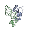
| Title | Structure of tmRNA-(SmpB)2 complex as inferred from cryo-EM | ||||||
 Components Components |
| ||||||
 Keywords Keywords | RNA BINDING PROTEIN/RNA / tmRNA / SmpB / RNA BINDING PROTEIN-RNA COMPLEX | ||||||
| Function / homology |  Function and homology information Function and homology information | ||||||
| Biological species |   Thermus thermophilus (bacteria) Thermus thermophilus (bacteria) | ||||||
| Method | ELECTRON MICROSCOPY / single particle reconstruction / cryo EM / Resolution: 13.6 Å | ||||||
 Authors Authors | Frank, J. / Felden, B. / Gillet, R. / Li, W. | ||||||
 Citation Citation |  Journal: J Biol Chem / Year: 2007 Journal: J Biol Chem / Year: 2007Title: Scaffolding as an organizing principle in trans-translation. The roles of small protein B and ribosomal protein S1. Authors: Reynald Gillet / Sukhjit Kaur / Wen Li / Marc Hallier / Brice Felden / Joachim Frank /  Abstract: A eubacterial ribosome stalled on a defective mRNA can be released through a quality control mechanism referred to as trans-translation, which depends on the coordinating binding actions of transfer- ...A eubacterial ribosome stalled on a defective mRNA can be released through a quality control mechanism referred to as trans-translation, which depends on the coordinating binding actions of transfer-messenger RNA, small protein B, and ribosome protein S1. By means of cryo-electron microscopy, we obtained a map of the complex composed of a stalled ribosome and small protein B, which appears near the decoding center. This result suggests that, when lacking a codon, the A-site on the small subunit is a target for small protein B. To investigate the role of S1 played in trans-translation, we obtained a cryo-electron microscopic map, including a stalled ribosome, transfer-messenger RNA, and small protein Bs but in the absence of S1. In this complex, several connections between the 30 S subunit and transfer-messenger RNA that appear in the +S1 complex are no longer found. We propose the unifying concept of scaffolding for the roles of small protein B and S1 in binding of transfer-messenger RNA to the ribosome during trans-translation, and we infer a pathway of sequential binding events in the initial phase of trans-translation. #1:  Journal: Proc.Natl.Acad.Sci.USA / Year: 2006 Journal: Proc.Natl.Acad.Sci.USA / Year: 2006Title: Cryo-EM visualization of transfer messenger RNA with two SmpBs in a stalled ribosome Authors: Kaur, S. / Gillet, R. / Li, W. / Gursky, R. / Frank, J. #2:  Journal: Science / Year: 2003 Journal: Science / Year: 2003Title: Visualizing tmRNA entry into a stalled ribosome Authors: Valle, M. / Gillet, R. / Kaur, K. / Henne, A. / Ramakrishnan, V. / Frank, J. | ||||||
| History |
| ||||||
| Remark 999 | SEQUENCE The deposited entry is a model to fit the cryo-EM map of tmRNA+SmpB(2) from Thermus ...SEQUENCE The deposited entry is a model to fit the cryo-EM map of tmRNA+SmpB(2) from Thermus thermophilus. The deposition includes 4 chains: chain A: tmRNA model chains B,C: SmpB (from 1P6V.pdb chain A, for SmpB from AQUIFEX AEOLICUS); chain D: helix 44 of 30S ribosomal subunit (from 1N34.pdb chain A:1406-1496. X-ray structure of 1N34 is from Thermus thermophilus). The E.coli model eschcolitm3D-model-72.pdb (http://www.ag.auburn.edu/mirror/tmRDB/rna/tmrna.html/) was used as a template to build the model by replacing several fragments which have x-ray crystal as alternatives, and by fitting all the structures into the cryo-EM maps. Nucleotide numbering in this model follows E.coli sequence. Considering the differences between tmRNA sequences from E.coli and T.thermophilus, a small number of nucleotides in the template model are not included in this model. |
- Structure visualization
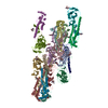
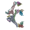
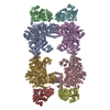
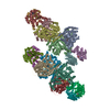
| Movie |
 Movie viewer Movie viewer |
|---|---|
| Structure viewer | Molecule:  Molmil Molmil Jmol/JSmol Jmol/JSmol |
- Downloads & links
- Download
| PDBx/mmCIF format |  2ob7.cif.gz 2ob7.cif.gz | 37.5 KB | Display |  PDBx/mmCIF format PDBx/mmCIF format |
|---|---|---|---|---|
| PDB format |  pdb2ob7.ent.gz pdb2ob7.ent.gz | 18.2 KB | Display |  PDB format PDB format |
| PDBx/mmJSON format |  2ob7.json.gz 2ob7.json.gz | Tree view |  PDBx/mmJSON format PDBx/mmJSON format | |
| Others |  Other downloads Other downloads |
-Validation report
| Arichive directory |  https://data.pdbj.org/pub/pdb/validation_reports/ob/2ob7 https://data.pdbj.org/pub/pdb/validation_reports/ob/2ob7 ftp://data.pdbj.org/pub/pdb/validation_reports/ob/2ob7 ftp://data.pdbj.org/pub/pdb/validation_reports/ob/2ob7 | HTTPS FTP |
|---|
-Related structure data
| Related structure data |  1310MC  1311M  1312C M: map data used to model this data C: citing same article ( |
|---|---|
| Similar structure data |
- Links
Links
- Assembly
| Deposited unit | 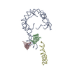
|
|---|---|
| 1 |
|
- Components
| #1: RNA chain | Mass: 105728.523 Da / Num. of mol.: 1 / Source method: isolated from a natural source / Source: (natural)   Thermus thermophilus (bacteria) Thermus thermophilus (bacteria) |
|---|---|
| #2: RNA chain | Mass: 27987.715 Da / Num. of mol.: 1 / Source method: isolated from a natural source / Details: helix 44 of 30S ribosomal subunit / Source: (natural)   Thermus thermophilus (bacteria) Thermus thermophilus (bacteria) |
| #3: Protein | Mass: 18440.818 Da / Num. of mol.: 2 / Source method: isolated from a natural source / Source: (natural)   Thermus thermophilus (bacteria) / References: UniProt: O66640*PLUS Thermus thermophilus (bacteria) / References: UniProt: O66640*PLUS |
-Experimental details
-Experiment
| Experiment | Method: ELECTRON MICROSCOPY |
|---|---|
| EM experiment | Aggregation state: PARTICLE / 3D reconstruction method: single particle reconstruction |
- Sample preparation
| Component | Name: Pre-accommodated ribosomal trans-translation complex: T. thermophilus 70S-mRNA-(P-site tRNA)-tmRNA-(SmpB)2-(EF-Tu)-GDP-kirromycin Type: RIBOSOME Details: see Experimental Procedures in Kaur et al. (PNAS). [Deposition refers to structure of tmRNA-(SmpB)2 complex derived by fitting of EM map from Kaur et al. Fitting was modified in Gillet et al.] |
|---|---|
| Buffer solution | Name: polimix / pH: 7.5 / Details: polimix |
| Specimen | Conc.: 32 mg/ml / Embedding applied: NO / Shadowing applied: NO / Staining applied: NO / Vitrification applied: YES |
| Specimen support | Details: This grid plus sample was kept at -80 degree C for several days before use. |
| Vitrification | Instrument: FEI VITROBOT MARK I / Cryogen name: ETHANE Details: Blot for 5 seconds before plunging. Rapid plunge freezing in liquid ethane. |
- Electron microscopy imaging
| Experimental equipment |  Model: Tecnai F20 / Image courtesy: FEI Company |
|---|---|
| Microscopy | Model: FEI TECNAI F20 / Date: Jun 1, 2004 |
| Electron gun | Electron source:  FIELD EMISSION GUN / Accelerating voltage: 200 kV / Illumination mode: FLOOD BEAM FIELD EMISSION GUN / Accelerating voltage: 200 kV / Illumination mode: FLOOD BEAM |
| Electron lens | Mode: BRIGHT FIELD / Nominal magnification: 55000 X / Calibrated magnification: 49000 X / Nominal defocus max: 3635 nm / Nominal defocus min: 1475 nm / Cs: 2 mm |
| Specimen holder | Temperature: 296 K / Tilt angle max: 0 ° / Tilt angle min: 0 ° |
| Image recording | Electron dose: 15 e/Å2 / Film or detector model: KODAK SO-163 FILM |
- Processing
| EM software |
| ||||||||||||
|---|---|---|---|---|---|---|---|---|---|---|---|---|---|
| CTF correction | Details: CTF correction of each defocus group reconstruction, by Wiener filtering | ||||||||||||
| Symmetry | Point symmetry: C1 (asymmetric) | ||||||||||||
| 3D reconstruction | Method: single particle reconstruction / Resolution: 13.6 Å / Num. of particles: 52829 / Nominal pixel size: 2.82 Å / Symmetry type: POINT | ||||||||||||
| Atomic model building | Protocol: OTHER / Space: REAL Details: METHOD--manual fitting using stereo visualization REFINEMENT PROTOCOL--Manual | ||||||||||||
| Atomic model building | PDB-ID: 1P6V Accession code: 1P6V / Details: 1P6V FOR SMPB AND TLD OF TMRNA / Source name: PDB / Type: experimental model | ||||||||||||
| Refinement step | Cycle: LAST
|
 Movie
Movie Controller




 PDBj
PDBj


