 Movie
Movie Controller

[English] 日本語
 Yorodumi
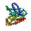
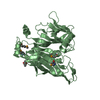
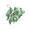
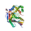
- PDB-1p6v: Crystal structure of the tRNA domain of transfer-messenger RNA in... -
+ Open data

- Basic information
| Entry | Database: PDB / ID: 1p6v | ||||||
|---|---|---|---|---|---|---|---|
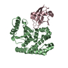
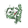
| Title | Crystal structure of the tRNA domain of transfer-messenger RNA in complex with SmpB | ||||||
 Components Components |
| ||||||
 Keywords Keywords | RNA BINDING PROTEIN/RNA / SmpB / tmRNA / trans-translation / protein-RNA complex / RNA BINDING PROTEIN-RNA COMPLEX | ||||||
| Function / homology |  Function and homology information Function and homology information | ||||||
| Biological species |   Aquifex aeolicus (bacteria) Aquifex aeolicus (bacteria) | ||||||
| Method |  X-RAY DIFFRACTION / SYNCHROTRON / MAD / Resolution: 3.2 Å X-RAY DIFFRACTION / SYNCHROTRON / MAD / Resolution: 3.2 Å | ||||||
 Authors Authors | Gutmann, S. / Haebel, P.W. / Metzinger, L. / Sutter, M. / Felden, B. / Ban, N. | ||||||
 Citation Citation | Journal: Nature / Year: 2003 Title: Crystal structure of the transfer-RNA domain of transfer-messenger RNA in complex with SmpB Authors: Gutmann, S. / Haebel, P.W. / Metzinger, L. / Sutter, M. / Felden, B. / Ban, N. | ||||||
| History |
|
- Structure visualization
| Structure viewer | Molecule: MolmilJmol/JSmol |
|---|
- Downloads & links
-Download
| PDBx/mmCIF format | 1p6v.cif.gz | 93.5 KB | Display | PDBx/mmCIF format |
|---|---|---|---|---|
| PDB format | pdb1p6v.ent.gz | 70 KB | Display | PDB format |
| PDBx/mmJSON format | 1p6v.json.gz | Tree view | PDBx/mmJSON format | |
| Others |  Other downloads Other downloads |
-Validation report
| Arichive directory | https://data.pdbj.org/pub/pdb/validation_reports/p6/1p6vftp://data.pdbj.org/pub/pdb/validation_reports/p6/1p6v | HTTPS FTP |
|---|
-Related structure data
| Similar structure data |
|---|


-Links
 PDBj
PDBj


- Assembly
| Deposited unit | 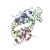
| ||||||||
|---|---|---|---|---|---|---|---|---|---|
| 1 | 
| ||||||||
| 2 | 
| ||||||||
| Unit cell |
| ||||||||
| Details | SmpB (chain A) and tmRNA[delta] (chain B) for the ribonucleoprotein complex / SmpB (chain C) and tmRNA[delta] (chain D) for the ribonucleoprotein complex; poorer defined in electron density |
-Components
| #1: RNA chain | Mass: 21959.070 Da / Num. of mol.: 2 / Source method: obtained synthetically Details: In vitro transcription of Aquifex aeolicus tmRNA[delta] #2: Protein | Mass: 18440.818 Da / Num. of mol.: 2 Source method: isolated from a genetically manipulated source Source: (gene. exp.) Aquifex aeolicus (bacteria) / Gene: SMPB / Production host: |
|---|
-Experimental details
-Experiment
| Experiment | Method: X-RAY DIFFRACTION / Number of used crystals: 1 |
|---|
- Sample preparation
| Crystal | Density Matthews: 5.22 Å3/Da / Density % sol: 76.25 % | |||||||||||||||||||||||||||||||||||||||||||||||||||||||||||||||||||||||||||||
|---|---|---|---|---|---|---|---|---|---|---|---|---|---|---|---|---|---|---|---|---|---|---|---|---|---|---|---|---|---|---|---|---|---|---|---|---|---|---|---|---|---|---|---|---|---|---|---|---|---|---|---|---|---|---|---|---|---|---|---|---|---|---|---|---|---|---|---|---|---|---|---|---|---|---|---|---|---|---|
| Crystal grow | Temperature: 293 K / Method: vapor diffusion, sitting drop / pH: 6.8 Details: Isopropanol, cacodylate, ammonium acetate, magnesium acetate, calcium acetate, pH 6.8, VAPOR DIFFUSION, SITTING DROP, temperature 293K | |||||||||||||||||||||||||||||||||||||||||||||||||||||||||||||||||||||||||||||
| Components of the solutions |
| |||||||||||||||||||||||||||||||||||||||||||||||||||||||||||||||||||||||||||||
| Crystal grow | *PLUS Temperature: 19 ℃ / Method: vapor diffusion, sitting drop | |||||||||||||||||||||||||||||||||||||||||||||||||||||||||||||||||||||||||||||
| Components of the solutions | *PLUS
|
-Data collection
| Diffraction | Mean temperature: 100 K |
|---|---|
| Diffraction source | Source: SYNCHROTRON / Site: SLS  / Beamline: X06SA / Wavelength: 1.008 Å / Beamline: X06SA / Wavelength: 1.008 Å |
| Detector | Type: MARRESEARCH / Detector: CCD / Date: Nov 12, 2002 |
| Radiation | Monochromator: Si 111 / Protocol: SINGLE WAVELENGTH / Monochromatic (M) / Laue (L): M / Scattering type: x-ray |
| Radiation wavelength | Wavelength: 1.008 Å / Relative weight: 1 |
| Reflection | Resolution: 3.2→50 Å / Num. all: 17756 / Num. obs: 17756 / % possible obs: 98.4 % / Observed criterion σ(F): 0 / Observed criterion σ(I): -3 / Redundancy: 6.9 % / Rsym value: 0.07 / Net I/σ(I): 23.1 |
| Reflection shell | Resolution: 3.2→3.3 Å / Redundancy: 7.3 % / Mean I/σ(I) obs: 2.4 / Rsym value: 0.87 / % possible all: 100 |
| Reflection | *PLUS Lowest resolution: 50 Å / Rmerge(I) obs: 0.075 |
| Reflection shell | *PLUS % possible obs: 100 % / Rmerge(I) obs: 0.873 |
- Processing
| Software |
| |||||||||||||||||||||||||
|---|---|---|---|---|---|---|---|---|---|---|---|---|---|---|---|---|---|---|---|---|---|---|---|---|---|---|
| Refinement | Method to determine structure: MAD / Resolution: 3.2→19.94 Å / Rfactor Rfree error: 0.009 / Isotropic thermal model: GROUPED B-Factors / Cross valid method: THROUGHOUT / σ(F): 0 / Stereochemistry target values: Engh & Huber
| |||||||||||||||||||||||||
| Solvent computation | Solvent model: FLAT MODEL / Bsol: 90.774 Å2 / ksol: 0.224974 e/Å3 | |||||||||||||||||||||||||
| Displacement parameters | Biso mean: 108.4 Å2
| |||||||||||||||||||||||||
| Refine analyze |
| |||||||||||||||||||||||||
| Refinement step | Cycle: LAST / Resolution: 3.2→19.94 Å
| |||||||||||||||||||||||||
| Refine LS restraints |
| |||||||||||||||||||||||||
| LS refinement shell | Resolution: 3.2→3.4 Å / Rfactor Rfree error: 0.032 / Total num. of bins used: 6
| |||||||||||||||||||||||||
| Xplor file |
| |||||||||||||||||||||||||
| Refinement | *PLUS Highest resolution: 3.2 Å / Lowest resolution: 20 Å / % reflection Rfree: 10 % / Rfactor Rfree: 0.36 | |||||||||||||||||||||||||
| Solvent computation | *PLUS | |||||||||||||||||||||||||
| Displacement parameters | *PLUS | |||||||||||||||||||||||||
| Refine LS restraints | *PLUS
|
