 Movie
Movie Controller


+ Open data

- Basic information
| Entry | Database: PDB / ID: 1pv6 | ||||||
|---|---|---|---|---|---|---|---|
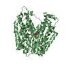
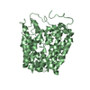
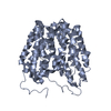
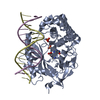
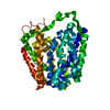
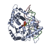
| Title | Crystal structure of lactose permease | ||||||
 Components Components | Lactose permease | ||||||
 Keywords Keywords | TRANSPORT PROTEIN / Transport / Sugar transport / Symport / membrane protein | ||||||
| Function / homology |  Function and homology information Function and homology informationlactose:proton symporter activity / lactose transport / carbohydrate:proton symporter activity / lactose binding / carbohydrate transport / membrane / plasma membrane Similarity search - Function | ||||||
| Biological species |  | ||||||
| Method |  X-RAY DIFFRACTION / SYNCHROTRON / MAD / Resolution: 3.5 Å X-RAY DIFFRACTION / SYNCHROTRON / MAD / Resolution: 3.5 Å | ||||||
 Authors Authors | Abramson, J. / Smirnova, I. / Kasho, V. / Verner, G. / Kaback, H.R. / Iwata, S. | ||||||
 Citation Citation | Journal: SCIENCE / Year: 2003 Title: Structure and mechanism of the lactose permease of Escherichia coli Authors: Abramson, J. / Smirnova, I. / Kasho, V. / Verner, G. / Kaback, H.R. / Iwata, S. | ||||||
| History |
|
- Structure visualization
| Structure viewer | Molecule: MolmilJmol/JSmol |
|---|
- Downloads & links
-Download
| PDBx/mmCIF format | 1pv6.cif.gz | 166.2 KB | Display | PDBx/mmCIF format |
|---|---|---|---|---|
| PDB format | pdb1pv6.ent.gz | 135.3 KB | Display | PDB format |
| PDBx/mmJSON format | 1pv6.json.gz | Tree view | PDBx/mmJSON format | |
| Others |  Other downloads Other downloads |
-Validation report
| Arichive directory | https://data.pdbj.org/pub/pdb/validation_reports/pv/1pv6ftp://data.pdbj.org/pub/pdb/validation_reports/pv/1pv6 | HTTPS FTP |
|---|
-Related structure data


-Links
 PDBj
PDBj


- Assembly
| Deposited unit | 
| ||||||||
|---|---|---|---|---|---|---|---|---|---|
| 1 | 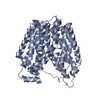
| ||||||||
| 2 | 
| ||||||||
| Unit cell |
|
-Components
| #1: Protein | Mass: 46484.797 Da / Num. of mol.: 2 / Mutation: C154G Source method: isolated from a genetically manipulated source Source: (gene. exp.) |
|---|
-Experimental details
-Experiment
| Experiment | Method: X-RAY DIFFRACTION / Number of used crystals: 1 |
|---|
- Sample preparation
| Crystal | Density Matthews: 6.45 Å3/Da | ||||||||||||||||||||||||||||||||||||||||||
|---|---|---|---|---|---|---|---|---|---|---|---|---|---|---|---|---|---|---|---|---|---|---|---|---|---|---|---|---|---|---|---|---|---|---|---|---|---|---|---|---|---|---|---|
| Crystal grow | Temperature: 293 K / Method: vapor diffusion, hanging drop / pH: 7 Details: PEG 400, pH 7.0, VAPOR DIFFUSION, HANGING DROP, temperature 293K | ||||||||||||||||||||||||||||||||||||||||||
| Crystal grow | *PLUS Temperature: 20 ℃ / Method: vapor diffusion, hanging drop | ||||||||||||||||||||||||||||||||||||||||||
| Components of the solutions | *PLUS
|
-Data collection
| Diffraction | Mean temperature: 100 K |
|---|---|
| Diffraction source | Source: SYNCHROTRON / Site: SLS  / Beamline: X06SA / Wavelength: 1.009 Å / Beamline: X06SA / Wavelength: 1.009 Å |
| Detector | Type: MARRESEARCH / Detector: CCD / Date: Mar 28, 2003 |
| Radiation | Monochromator: Si 111 / Protocol: MAD / Monochromatic (M) / Laue (L): M / Scattering type: x-ray |
| Radiation wavelength | Wavelength: 1.009 Å / Relative weight: 1 |
| Reflection | Resolution: 3.5→40 Å / Num. all: 31004 / Num. obs: 25733 / % possible obs: 83 % / Observed criterion σ(F): 0 / Observed criterion σ(I): -1 |
| Reflection shell | Resolution: 3.5→3.6 Å / % possible all: 71.8 |
| Reflection | *PLUS Num. obs: 25917 / % possible obs: 85.9 % / Redundancy: 2.9 % / Num. measured all: 63863 / Rmerge(I) obs: 0.075 |
| Reflection shell | *PLUS % possible obs: 71.8 % / Rmerge(I) obs: 0.461 |
- Processing
| Software |
| ||||||||||||||||||||
|---|---|---|---|---|---|---|---|---|---|---|---|---|---|---|---|---|---|---|---|---|---|
| Refinement | Method to determine structure: MAD / Resolution: 3.5→40 Å / σ(F): 0 / Stereochemistry target values: Engh & Huber
| ||||||||||||||||||||
| Refinement step | Cycle: LAST / Resolution: 3.5→40 Å
| ||||||||||||||||||||
| Refine LS restraints |
| ||||||||||||||||||||
| Refinement | *PLUS Lowest resolution: 40 Å / % reflection Rfree: 5 % | ||||||||||||||||||||
| Solvent computation | *PLUS | ||||||||||||||||||||
| Displacement parameters | *PLUS | ||||||||||||||||||||
| Refine LS restraints | *PLUS
| ||||||||||||||||||||
| LS refinement shell | *PLUS Highest resolution: 3.5 Å / Lowest resolution: 3.6 Å / Rfactor Rfree: 0.379 / Rfactor Rwork: 0.356 |
