 Movie
Movie Controller


+ Open data

- Basic information
| Entry | Database: PDB / ID: 2zur | ||||||
|---|---|---|---|---|---|---|---|
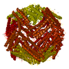
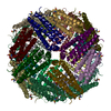
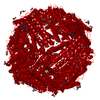
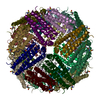
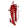
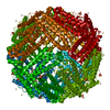
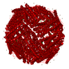
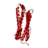
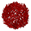
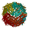
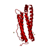
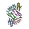
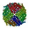
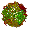
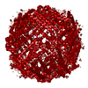
| Title | Crystal Structure of Rh(nbd)/apo-Fr | ||||||
 Components Components | Ferritin light chain | ||||||
 Keywords Keywords | METAL BINDING PROTEIN / Acetylation / Iron / Iron storage / Metal-binding | ||||||
| Function / homology |  Function and homology information Function and homology informationferritin complex / autolysosome / ferric iron binding / autophagosome / ferrous iron binding / iron ion transport / cytoplasmic vesicle / intracellular iron ion homeostasis / iron ion binding / cytoplasm Similarity search - Function | ||||||
| Biological species |  | ||||||
| Method |  X-RAY DIFFRACTION / SYNCHROTRON / MOLECULAR REPLACEMENT / Resolution: 1.8 Å X-RAY DIFFRACTION / SYNCHROTRON / MOLECULAR REPLACEMENT / Resolution: 1.8 Å | ||||||
 Authors Authors | Abe, S. / Hirata, K. / Ueno, T. / Shimizu, N. / Yamamoto, M. / Takata, M. / Watanabe, Y. | ||||||
 Citation Citation | Journal: J.Am.Chem.Soc. / Year: 2009 Title: Polymerization of phenylacetylene by rhodium complexes within a discrete space of apo-ferritin Authors: Abe, S. / Hirata, K. / Ueno, T. / Morino, K. / Shimizu, N. / Yamamoto, M. / Takata, M. / Yashima, E. / Watanabe, Y. | ||||||
| History |
| ||||||
| Remark 650 | HELIX DETERMINATION METHOD: AUTHOR DETERMINED |
- Structure visualization
| Structure viewer | Molecule: MolmilJmol/JSmol |
|---|
- Downloads & links
-Download
| PDBx/mmCIF format | 2zur.cif.gz | 58.3 KB | Display | PDBx/mmCIF format |
|---|---|---|---|---|
| PDB format | pdb2zur.ent.gz | 43.4 KB | Display | PDB format |
| PDBx/mmJSON format | 2zur.json.gz | Tree view | PDBx/mmJSON format | |
| Others |  Other downloads Other downloads |
-Validation report
| Summary document | 2zur_validation.pdf.gz | 462.6 KB | Display | wwPDB validaton report |
|---|---|---|---|---|
| Full document | 2zur_full_validation.pdf.gz | 464.4 KB | Display | |
| Data in XML | 2zur_validation.xml.gz | 12 KB | Display | |
| Data in CIF | 2zur_validation.cif.gz | 17 KB | Display | |
| Arichive directory | https://data.pdbj.org/pub/pdb/validation_reports/zu/2zurftp://data.pdbj.org/pub/pdb/validation_reports/zu/2zur | HTTPS FTP |
-Related structure data
| Related structure data |  1datS S: Starting model for refinement |
|---|---|
| Similar structure data |

-Links
 PDBj
PDBj


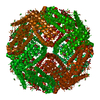
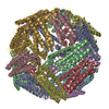
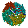
- Assembly
| Deposited unit | 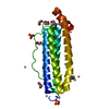
| |||||||||||||||
|---|---|---|---|---|---|---|---|---|---|---|---|---|---|---|---|---|
| 1 | x 24
| |||||||||||||||
| Unit cell |
| |||||||||||||||
| Components on special symmetry positions |
|
-Components
-Protein , 1 types, 1 molecules X
| #1: Protein | Mass: 20066.465 Da / Num. of mol.: 1 Source method: isolated from a genetically manipulated source Source: (gene. exp.)  |
|---|
-Non-polymers , 5 types, 209 molecules 


| #2: Chemical | ChemComp-CD /  Mass: 112.411 Da / Num. of mol.: 5 / Source method: obtained synthetically / Formula: Cd Mass: 112.411 Da / Num. of mol.: 5 / Source method: obtained synthetically / Formula: Cd#3: Chemical |  Mass: 96.063 Da / Num. of mol.: 2 / Source method: obtained synthetically / Formula: SO4 Mass: 96.063 Da / Num. of mol.: 2 / Source method: obtained synthetically / Formula: SO4#4: Chemical |  Mass: 102.906 Da / Num. of mol.: 2 / Source method: obtained synthetically / Formula: Rh Mass: 102.906 Da / Num. of mol.: 2 / Source method: obtained synthetically / Formula: Rh#5: Chemical | ChemComp-EDO /  Mass: 62.068 Da / Num. of mol.: 8 / Source method: obtained synthetically / Formula: C2H6O2 Mass: 62.068 Da / Num. of mol.: 8 / Source method: obtained synthetically / Formula: C2H6O2#6: Water | ChemComp-HOH / | Mass: 18.015 Da / Num. of mol.: 192 / Source method: isolated from a natural source / Formula: H2O |
|---|
-Details
| Has protein modification | Y |
|---|
-Experimental details
-Experiment
| Experiment | Method: X-RAY DIFFRACTION / Number of used crystals: 1 |
|---|
- Sample preparation
| Crystal | Density Matthews: 3.09 Å3/Da / Density % sol: 60.22 % |
|---|---|
| Crystal grow | Temperature: 293 K / Method: vapor diffusion, hanging drop / pH: 7 Details: 0.5M ammonium sulfate, 10mM cadmium sulfate, pH 7, VAPOR DIFFUSION, HANGING DROP, temperature 293K |
-Data collection
| Diffraction | Mean temperature: 100 K |
|---|---|
| Diffraction source | Source: SYNCHROTRON / Site: SPring-8  / Beamline: BL41XU / Wavelength: 0.5334 Å / Beamline: BL41XU / Wavelength: 0.5334 Å |
| Detector | Type: ADSC QUANTUM 315 / Detector: CCD / Date: Mar 3, 2006 |
| Radiation | Monochromator: Si 111 Channel / Protocol: SINGLE WAVELENGTH / Monochromatic (M) / Laue (L): M / Scattering type: x-ray |
| Radiation wavelength | Wavelength: 0.5334 Å / Relative weight: 1 |
| Reflection | Resolution: 1.8→50 Å / Num. obs: 24236 / % possible obs: 100 % / Redundancy: 11.6 % / Biso Wilson estimate: 18.6 Å2 / Rmerge(I) obs: 0.073 / Net I/σ(I): 38.2 |
| Reflection shell | Resolution: 1.8→1.86 Å / Redundancy: 11.7 % / Rmerge(I) obs: 0.327 / Mean I/σ(I) obs: 9.3 / % possible all: 100 |
- Processing
| Software |
| ||||||||||||||||||||||||||||||||||||||||||||||||||||||||||||||||||||||||||||||||||||||||||||||||||||
|---|---|---|---|---|---|---|---|---|---|---|---|---|---|---|---|---|---|---|---|---|---|---|---|---|---|---|---|---|---|---|---|---|---|---|---|---|---|---|---|---|---|---|---|---|---|---|---|---|---|---|---|---|---|---|---|---|---|---|---|---|---|---|---|---|---|---|---|---|---|---|---|---|---|---|---|---|---|---|---|---|---|---|---|---|---|---|---|---|---|---|---|---|---|---|---|---|---|---|---|---|---|
| Refinement | Method to determine structure: MOLECULAR REPLACEMENT Starting model: PDB ENTRY 1DAT Resolution: 1.8→37.01 Å / Cor.coef. Fo:Fc: 0.95 / Cor.coef. Fo:Fc free: 0.947 / SU B: 1.684 / SU ML: 0.055 / Cross valid method: THROUGHOUT / ESU R: 0.105 / ESU R Free: 0.098 / Stereochemistry target values: MAXIMUM LIKELIHOOD
| ||||||||||||||||||||||||||||||||||||||||||||||||||||||||||||||||||||||||||||||||||||||||||||||||||||
| Solvent computation | Ion probe radii: 0.8 Å / Shrinkage radii: 0.8 Å / VDW probe radii: 1.2 Å / Solvent model: MASK | ||||||||||||||||||||||||||||||||||||||||||||||||||||||||||||||||||||||||||||||||||||||||||||||||||||
| Displacement parameters | Biso mean: 19.801 Å2 | ||||||||||||||||||||||||||||||||||||||||||||||||||||||||||||||||||||||||||||||||||||||||||||||||||||
| Refinement step | Cycle: LAST / Resolution: 1.8→37.01 Å
| ||||||||||||||||||||||||||||||||||||||||||||||||||||||||||||||||||||||||||||||||||||||||||||||||||||
| Refine LS restraints |
| ||||||||||||||||||||||||||||||||||||||||||||||||||||||||||||||||||||||||||||||||||||||||||||||||||||
| LS refinement shell | Resolution: 1.801→1.847 Å / Total num. of bins used: 20
|
