[English] 日本語
 Yorodumi
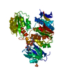
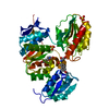
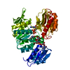
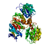
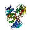
- PDB-2vtd: Crystal structure of MurD ligase in complex with D-Glu containing... -
+ Open data
- Basic information
| Entry | Database: PDB / ID: 2vtd | ||||||
|---|---|---|---|---|---|---|---|
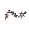
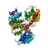
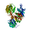
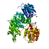
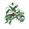
| Title | Crystal structure of MurD ligase in complex with D-Glu containing sulfonamide inhibitor | ||||||
 Components Components | UDP-N-ACETYLMURAMOYLALANINE--D-GLUTAMATE LIGASE | ||||||
 Keywords Keywords | LIGASE / MURD-INHIBITOR COMPLEX / PEPTIDOGLYCAN SYNTHESIS / NUCLEOTIDE-BINDING / SULFONAMIDE INHIBITOR / MURD LIGASE / ATP-BINDING / CELL DIVISION / CYTOPLASM / CELL SHAPE / CELL CYCLE / CELL WALL BIOGENESIS/DEGRADATION | ||||||
| Function / homology |  Function and homology information Function and homology informationUDP-N-acetylmuramoyl-L-alanine-D-glutamate ligase / UDP-N-acetylmuramoylalanine-D-glutamate ligase activity / peptidoglycan biosynthetic process / cell wall organization / regulation of cell shape / cell division / ATP binding / identical protein binding / cytoplasm Similarity search - Function | ||||||
| Biological species |  | ||||||
| Method |  X-RAY DIFFRACTION / X-RAY DIFFRACTION /  SYNCHROTRON / SYNCHROTRON /  MOLECULAR REPLACEMENT / Resolution: 1.94 Å MOLECULAR REPLACEMENT / Resolution: 1.94 Å | ||||||
 Authors Authors | Humljan, J. / Kotnik, M. / Contreras-Martel, C. / Blanot, D. / Urleb, U. / Dessen, A. / Solmajer, T. / Gobec, S. | ||||||
 Citation Citation |  Journal: J. Med. Chem. / Year: 2008 Journal: J. Med. Chem. / Year: 2008Title: Novel naphthalene-N-sulfonyl-D-glutamic acid derivatives as inhibitors of MurD, a key peptidoglycan biosynthesis enzyme. Authors: Humljan, J. / Kotnik, M. / Contreras-Martel, C. / Blanot, D. / Urleb, U. / Dessen, A. / Solmajer, T. / Gobec, S. | ||||||
| History |
|
- Structure visualization
| Structure viewer | Molecule:  Molmil Molmil Jmol/JSmol Jmol/JSmol |
|---|
- Downloads & links
- Download
| PDBx/mmCIF format |  2vtd.cif.gz 2vtd.cif.gz | 105 KB | Display |  PDBx/mmCIF format PDBx/mmCIF format |
|---|---|---|---|---|
| PDB format |  pdb2vtd.ent.gz pdb2vtd.ent.gz | 77.3 KB | Display |  PDB format PDB format |
| PDBx/mmJSON format |  2vtd.json.gz 2vtd.json.gz | Tree view |  PDBx/mmJSON format PDBx/mmJSON format | |
| Others |  Other downloads Other downloads |
-Validation report
| Summary document |  2vtd_validation.pdf.gz 2vtd_validation.pdf.gz | 730.3 KB | Display |  wwPDB validaton report wwPDB validaton report |
|---|---|---|---|---|
| Full document |  2vtd_full_validation.pdf.gz 2vtd_full_validation.pdf.gz | 734.5 KB | Display | |
| Data in XML |  2vtd_validation.xml.gz 2vtd_validation.xml.gz | 20.9 KB | Display | |
| Data in CIF |  2vtd_validation.cif.gz 2vtd_validation.cif.gz | 31.2 KB | Display | |
| Arichive directory |  https://data.pdbj.org/pub/pdb/validation_reports/vt/2vtd https://data.pdbj.org/pub/pdb/validation_reports/vt/2vtd ftp://data.pdbj.org/pub/pdb/validation_reports/vt/2vtd ftp://data.pdbj.org/pub/pdb/validation_reports/vt/2vtd | HTTPS FTP |
-Related structure data
| Related structure data | 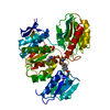 2uuoC 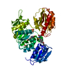 2uupC 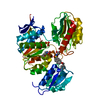 2vteC 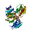 3uagS C: citing same article ( S: Starting model for refinement |
|---|---|
| Similar structure data |
- Links
Links
- Assembly
| Deposited unit | 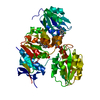
| ||||||||
|---|---|---|---|---|---|---|---|---|---|
| 1 |
| ||||||||
| Unit cell |
|
- Components
| #1: Protein | Mass: 47979.398 Da / Num. of mol.: 1 Source method: isolated from a genetically manipulated source Details: INHIBITOR N-(6-(4-CYANO-2-FLUORO-BENZYLOXY)-NAPHTHALENE-2-SULFONYL)-D-GLUTAMIC ACID BOUND Source: (gene. exp.)   References: UniProt: P14900, UDP-N-acetylmuramoyl-L-alanine-D-glutamate ligase |
|---|---|
| #2: Chemical | ChemComp-LKM / |
| #3: Chemical | ChemComp-SO4 / |
| #4: Water | ChemComp-HOH / |
| Sequence details | C-TERMINAL HIS-TAG (SHHHHHH) |
-Experimental details
-Experiment
| Experiment | Method:  X-RAY DIFFRACTION / Number of used crystals: 1 X-RAY DIFFRACTION / Number of used crystals: 1 |
|---|
- Sample preparation
| Crystal | Density Matthews: 3 Å3/Da / Density % sol: 59 % / Description: NONE |
|---|---|
| Crystal grow | pH: 7.5 Details: PROTEIN WAS CRYSTALLIZED FROM 1.7 M (NH4)2SO4, 7% PEG 400, 100 MM HEPES, PH 7.5; THEN SOAKED IN 2 MM OF INHIBITOR SOLUTION |
-Data collection
| Diffraction | Mean temperature: 287 K |
|---|---|
| Diffraction source | Source:  SYNCHROTRON / Site: SYNCHROTRON / Site:  ESRF ESRF  / Beamline: ID29 / Wavelength: 0.97623 / Beamline: ID29 / Wavelength: 0.97623 |
| Detector | Type: ADSC CCD / Detector: CCD / Date: Apr 30, 2008 |
| Radiation | Protocol: SINGLE WAVELENGTH / Monochromatic (M) / Laue (L): M / Scattering type: x-ray |
| Radiation wavelength | Wavelength: 0.97623 Å / Relative weight: 1 |
| Reflection | Resolution: 1.94→50 Å / Num. obs: 39330 / % possible obs: 94.5 % / Observed criterion σ(I): 3 / Redundancy: 4.6 % / Rmerge(I) obs: 0.05 / Net I/σ(I): 24 |
| Reflection shell | Resolution: 1.94→2.06 Å / Redundancy: 4.7 % / Rmerge(I) obs: 0.37 / Mean I/σ(I) obs: 5 / % possible all: 88 |
- Processing
| Software |
| ||||||||||||||||||||||||||||||||||||||||||||||||||||||||||||||||||||||||||||||||||||||||||||||||||||||||||||||||||||||||||||||||||||||||||||||||||||||||||||||||||||||||||||||||||||||
|---|---|---|---|---|---|---|---|---|---|---|---|---|---|---|---|---|---|---|---|---|---|---|---|---|---|---|---|---|---|---|---|---|---|---|---|---|---|---|---|---|---|---|---|---|---|---|---|---|---|---|---|---|---|---|---|---|---|---|---|---|---|---|---|---|---|---|---|---|---|---|---|---|---|---|---|---|---|---|---|---|---|---|---|---|---|---|---|---|---|---|---|---|---|---|---|---|---|---|---|---|---|---|---|---|---|---|---|---|---|---|---|---|---|---|---|---|---|---|---|---|---|---|---|---|---|---|---|---|---|---|---|---|---|---|---|---|---|---|---|---|---|---|---|---|---|---|---|---|---|---|---|---|---|---|---|---|---|---|---|---|---|---|---|---|---|---|---|---|---|---|---|---|---|---|---|---|---|---|---|---|---|---|---|
| Refinement | Method to determine structure:  MOLECULAR REPLACEMENT MOLECULAR REPLACEMENTStarting model: PDB ENTRY 3UAG Resolution: 1.94→46.88 Å / Cor.coef. Fo:Fc: 0.946 / Cor.coef. Fo:Fc free: 0.927 / Cross valid method: THROUGHOUT / ESU R: 0.145 / ESU R Free: 0.137 / Stereochemistry target values: MAXIMUM LIKELIHOOD Details: HYDROGENS HAVE BEEN ADDED IN THE RIDING POSITIONS. THE FOLLOWING RESIDUES WERE NOT LOCATED IN THE EXPERIMENT - - GLY A 222, ALA A 223, ASP A 224, GLN A 242, GLN A 243. RESIDUES ARG A 221 AND ...Details: HYDROGENS HAVE BEEN ADDED IN THE RIDING POSITIONS. THE FOLLOWING RESIDUES WERE NOT LOCATED IN THE EXPERIMENT - - GLY A 222, ALA A 223, ASP A 224, GLN A 242, GLN A 243. RESIDUES ARG A 221 AND HIS A 241 WERE MODELED AS ALANINES DUE TO INSUFFICIENT ELECTRON DENSITY.
| ||||||||||||||||||||||||||||||||||||||||||||||||||||||||||||||||||||||||||||||||||||||||||||||||||||||||||||||||||||||||||||||||||||||||||||||||||||||||||||||||||||||||||||||||||||||
| Solvent computation | Ion probe radii: 0.8 Å / Shrinkage radii: 0.8 Å / VDW probe radii: 1.4 Å / Solvent model: MASK | ||||||||||||||||||||||||||||||||||||||||||||||||||||||||||||||||||||||||||||||||||||||||||||||||||||||||||||||||||||||||||||||||||||||||||||||||||||||||||||||||||||||||||||||||||||||
| Displacement parameters | Biso mean: 24.01 Å2
| ||||||||||||||||||||||||||||||||||||||||||||||||||||||||||||||||||||||||||||||||||||||||||||||||||||||||||||||||||||||||||||||||||||||||||||||||||||||||||||||||||||||||||||||||||||||
| Refinement step | Cycle: LAST / Resolution: 1.94→46.88 Å
| ||||||||||||||||||||||||||||||||||||||||||||||||||||||||||||||||||||||||||||||||||||||||||||||||||||||||||||||||||||||||||||||||||||||||||||||||||||||||||||||||||||||||||||||||||||||
| Refine LS restraints |
|
 Movie
Movie Controller








 PDBj
PDBj