Mass: 18.015 Da / Num. of mol.: 296 / Source method: isolated from a natural source / Formula: H2O
Has protein modification
Y
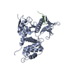
Sequence details
THE FIRST 30 RESIDUES CORRESPOND TO A SIGNAL PEPTIDE THAT IS PREDICTED TO HAVE CLEAVED DURING ...THE FIRST 30 RESIDUES CORRESPOND TO A SIGNAL PEPTIDE THAT IS PREDICTED TO HAVE CLEAVED DURING EXPRESSION OF THE PROTEIN.
-
Experimental details
-
Experiment
Experiment
Method: X-RAY DIFFRACTION / Number of used crystals: 1
-
Sample preparation
Crystal
Density Matthews: 2.2 Å3/Da / Density % sol: 43.2 %
Crystal grow
pH: 6.5 Details: 15 MG/ML PROTEIN. 25% PEG3350 AND 0.1 M BIS-TRIS HCL, PH 6.5.
Monochromator: DOUBLE CRYSTAL, SI(111) OR SI(311) / Protocol: SINGLE WAVELENGTH / Monochromatic (M) / Laue (L): M / Scattering type: x-ray
Radiation wavelength
Wavelength: 0.97931 Å / Relative weight: 1
Reflection
Resolution: 2.2→30 Å / Num. obs: 27378 / % possible obs: 99.9 % / Redundancy: 6 % / Rmerge(I) obs: 0.11 / Net I/σ(I): 13.7
Reflection shell
Resolution: 2.2→2.32 Å / Redundancy: 6.1 % / Rmerge(I) obs: 0.4 / Mean I/σ(I) obs: 4.7 / % possible all: 100
-
Processing
Software
Name
Version
Classification
REFMAC
5.2.0019
refinement
MOSFLM
datareduction
SCALA
datascaling
autoSHARP
phasing
Refinement
Method to determine structure: SAD / Resolution: 2.2→28.57 Å / Cor.coef. Fo:Fc: 0.949 / Cor.coef. Fo:Fc free: 0.92 / SU B: 4.672 / SU ML: 0.122 / Cross valid method: THROUGHOUT / ESU R: 0.259 / ESU R Free: 0.196 / Stereochemistry target values: MAXIMUM LIKELIHOOD Details: HYDROGENS HAVE BEEN ADDED IN THE RIDING POSITIONS. NO ELECTRON DENSITY CAN BE OBSERVED FOR THE CHAIN BETWEEN 101 AND 113.
Rfactor
Num. reflection
% reflection
Selection details
Rfree
0.214
1384
5.1 %
RANDOM
Rwork
0.163
-
-
-
obs
0.166
25985
100 %
-
Solvent computation
Ion probe radii: 0.8 Å / Shrinkage radii: 0.8 Å / VDW probe radii: 1.2 Å / Solvent model: MASK
 Movie
Movie Controller

 Yorodumi
 Open data
 Basic information
 Components
 Keywords
 Function and homology information
 X-RAY DIFFRACTION /
  Authors
 Citation
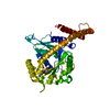
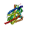
 Structure visualization
 Downloads & links
 Other downloads


 PDBj
PDBj
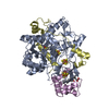
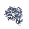
 Assembly
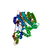
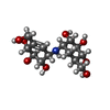
 Mass: 335.350 Da / Num. of mol.: 1 / Source method: obtained synthetically / Formula: C14H25NO8
 Mass: 18.015 Da / Num. of mol.: 296 / Source method: isolated from a natural source / Formula: H2O
 Sample preparation
 / Beamline: ID14-4 / Wavelength: 0.97931
  Processing