- PDB-1e08: Structural model of the [Fe]-Hydrogenase/cytochrome c553 complex ... -
+
Open data
ID or keywords:
Loading...
-
Basic information
Entry
Database: PDB / ID: 100000000
Title
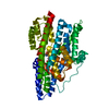
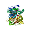
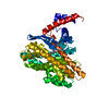
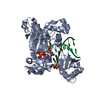
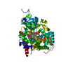
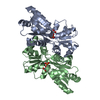
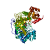
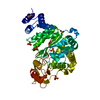
Structural model of the [Fe]-Hydrogenase/cytochrome c553 complex combining NMR and soft-docking
Components
([FE]-HYDROGENASE ...) x 2
CYTOCHROME C553
Keywords
HYDROGENASE / CYTOCHROME C553 / ELECTRON TRANSFER COMPLEX
Function / homology
Function and homology information
ferredoxin hydrogenase / ferredoxin hydrogenase activity / iron-sulfur cluster binding / 4 iron, 4 sulfur cluster binding / periplasmic space / electron transfer activity / iron ion binding / heme binding / metal ion binding Similarity search - Function
Iron hydrogenase, small subunit HydB-type / Iron hydrogenase, small subunit superfamily / Iron hydrogenase, subset / Iron hydrogenase, small subunit / : / Iron hydrogenase small subunit / Iron hydrogenase small subunit / Iron hydrogenase, large subunit, C-terminal / Iron hydrogenase / Iron only hydrogenase large subunit, C-terminal domain ...Iron hydrogenase, small subunit HydB-type / Iron hydrogenase, small subunit superfamily / Iron hydrogenase, subset / Iron hydrogenase, small subunit / : / Iron hydrogenase small subunit / Iron hydrogenase small subunit / Iron hydrogenase, large subunit, C-terminal / Iron hydrogenase / Iron only hydrogenase large subunit, C-terminal domain / 4Fe-4S dicluster domain / 4Fe-4S binding domain / Twin-arginine translocation pathway, signal sequence, bacterial/archaeal / Cytochrome c / Cytochrome c family profile. / Cytochrome c-like domain / Cytochrome c-like domain superfamily / Twin arginine translocation (Tat) signal profile. / Twin-arginine translocation pathway, signal sequence / 4Fe-4S ferredoxin, iron-sulphur binding, conserved site / 4Fe-4S ferredoxin-type iron-sulfur binding region signature. / 4Fe-4S ferredoxin-type iron-sulfur binding domain profile. / 4Fe-4S ferredoxin-type, iron-sulphur binding domain Similarity search - Domain/homology
CARBON MONOXIDE / CYANIDE ION / : / HEME C / 1,3-PROPANEDITHIOL / IRON/SULFUR CLUSTER / Cytochrome c-553 / Periplasmic [Fe] hydrogenase large subunit / Periplasmic [Fe] hydrogenase small subunit Similarity search - Component
Mass: 18.015 Da / Num. of mol.: 1 / Source method: isolated from a natural source / Formula: H2O
-
Details
Has protein modification
Y
Sequence details
N-TERMINAL END C-TERMINAL RESIDUES WERE NOT DEPOSITED IN THE INITIAL PDB ENTRY 1HFE TER ALA: THE C- ...N-TERMINAL END C-TERMINAL RESIDUES WERE NOT DEPOSITED IN THE INITIAL PDB ENTRY 1HFE TER ALA: THE C-TERMINAL RESIDUES 398-421 ARE NOT PRESENT IN THE PDB FILE OF HYDROGENASE 1HFE
-
Experimental details
-
Experiment
Experiment
Method
SOLUTION NMR
THEORETICAL MODEL
NMR experiment
Type: TROSY
NMR details
Text: COMBINED DOCKING AND NMR FILTERING SOLUTION. THE TROSY EXPERIMENT WAS PERFORMED ON THE 15N-LABELED CYTOCHROME WITH SUBSEQUENTLY ADDING OF THE HYDROGENASE. THE RESIDUES HAVING THE STRONGEST ...Text: COMBINED DOCKING AND NMR FILTERING SOLUTION. THE TROSY EXPERIMENT WAS PERFORMED ON THE 15N-LABELED CYTOCHROME WITH SUBSEQUENTLY ADDING OF THE HYDROGENASE. THE RESIDUES HAVING THE STRONGEST SHIFTS WERE USED TO FILTER THE DOCKING SOLUTIONS.THE AMINO-ACIDS ALA E5, CYS E10, HIS E14, GLY E15, ALA E16, ALA E22, GLY E24, VAL E29, GLN E32, LYS E54, ASN E59, ALA E60 OF THE CYTOCHROME C553 SHOW STRONGEST SHIFTS IN THE TROSY EXPERIMENTS. EACH TIME ONE OF THE AMINO ACIDS IS IN CONTACT (LESS THAN 5A IN THE COMPLEX) WITH ITS PARTNER ONE POINT IS ATTRIBUTED TO THE STRUCTURE. THEN THE ONE THOUSAND BEST STRUCTURES ARE RANKED ACCORDING TO THIS CRITERIUM AND THE 10 BEST SOLUTIONS ARE MINIMISED. A COMPARISON OF THESE SOLUTIONS GENERATES STRUCTURE WITH 5 IDENTICAL SOLUTIONS
Type: Bruker DRX / Manufacturer: Bruker / Model: DRX / Field strength: 500 MHz
-
Processing
NMR software
Name
Version
Developer
Classification
X-PLOR
3.843
BRUNGER
refinement
UXNMR
UXNMR
structuresolution
Refinement
Software ordinal: 1 Details: THE DEPOSITED STRUCTURE IS THE RESULT OF A HETERONUCLEAR NMR EXPERIMENT AND DOCKING SIMULATIONS. THE FINAL RESULT WAS MINIMIZED BY MOLECULAR DYNAMIC MINIMIZATION.
NMR ensemble
Conformer selection criteria: LEAST SHIFT VARIATION VIOLATION Conformers calculated total number: 1000 / Conformers submitted total number: 1
+
About Yorodumi
-
News
-
Feb 9, 2022. New format data for meta-information of EMDB entries
New format data for meta-information of EMDB entries
Version 3 of the EMDB header file is now the official format.
The previous official version 1.9 will be removed from the archive.
In the structure databanks used in Yorodumi, some data are registered as the other names, "COVID-19 virus" and "2019-nCoV". Here are the details of the virus and the list of structure data.
Jan 31, 2019. EMDB accession codes are about to change! (news from PDBe EMDB page)
EMDB accession codes are about to change! (news from PDBe EMDB page)
The allocation of 4 digits for EMDB accession codes will soon come to an end. Whilst these codes will remain in use, new EMDB accession codes will include an additional digit and will expand incrementally as the available range of codes is exhausted. The current 4-digit format prefixed with “EMD-” (i.e. EMD-XXXX) will advance to a 5-digit format (i.e. EMD-XXXXX), and so on. It is currently estimated that the 4-digit codes will be depleted around Spring 2019, at which point the 5-digit format will come into force.
The EM Navigator/Yorodumi systems omit the EMD- prefix.
Related info.:Q: What is EMD? / ID/Accession-code notation in Yorodumi/EM Navigator
Yorodumi is a browser for structure data from EMDB, PDB, SASBDB, etc.
This page is also the successor to EM Navigator detail page, and also detail information page/front-end page for Omokage search.
The word "yorodu" (or yorozu) is an old Japanese word meaning "ten thousand". "mi" (miru) is to see.
Related info.:EMDB / PDB / SASBDB / Comparison of 3 databanks / Yorodumi Search / Aug 31, 2016. New EM Navigator & Yorodumi / Yorodumi Papers / Jmol/JSmol / Function and homology information / Changes in new EM Navigator and Yorodumi
 Movie
Movie Controller

 Yorodumi
 Open data
 Basic information
 Components
 Keywords
 Function and homology information
 DESULFOVIBRIO DESULFURICANS (bacteria)
 Authors
 Citation
 Structure visualization
 Downloads & links
 Other downloads
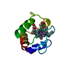


 PDBj
PDBj


 Assembly


 Mass: 351.640 Da / Num. of mol.: 3 / Source method: obtained synthetically / Formula: Fe4S4
 Mass: 108.226 Da / Num. of mol.: 1 / Source method: obtained synthetically / Formula: C3H8S2
 Mass: 55.845 Da / Num. of mol.: 2 / Source method: obtained synthetically / Formula: Fe
 Mass: 26.017 Da / Num. of mol.: 2 / Source method: obtained synthetically / Formula: CN
 Mass: 28.010 Da / Num. of mol.: 2 / Source method: obtained synthetically / Formula: CO
 Mass: 65.409 Da / Num. of mol.: 1 / Source method: obtained synthetically / Formula: Zn
 Mass: 618.503 Da / Num. of mol.: 1 / Source method: obtained synthetically / Formula: C34H34FeN4O4
 Sample preparation
 Processing
 X-PLOR