 Movie
Movie Controller


+ Open data

- Basic information
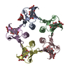
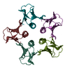
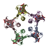
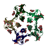
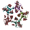
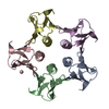
| Entry | Database: PDB / ID: 2bos | |||||||||
|---|---|---|---|---|---|---|---|---|---|---|
| Title | A MUTANT SHIGA-LIKE TOXIN IIE BOUND TO ITS RECEPTOR | |||||||||
 Components Components | PROTEIN (SHIGA-LIKE TOXIN IIE B SUBUNIT) | |||||||||
 Keywords Keywords | TOXIN / RECEPTOR BINDING / PROTEIN-CARBOHYDRATE RECOGNITION / SPECIFICITY | |||||||||
| Function / homology |  Function and homology information Function and homology informationsymbiont-mediated hemolysis of host erythrocyte / toxin activity / extracellular region Similarity search - Function | |||||||||
| Biological species |  | |||||||||
| Method |  X-RAY DIFFRACTION / MOLECULAR REPLACEMENT / Resolution: 2 Å X-RAY DIFFRACTION / MOLECULAR REPLACEMENT / Resolution: 2 Å | |||||||||
 Authors Authors | Ling, H. / Boodhoo, A. / Armstrong, G.D. / Clark, C.G. / Brunton, J.L. / Read, R.J. | |||||||||
 Citation Citation | Journal: Structure Fold.Des. / Year: 2000 Title: A mutant Shiga-like toxin IIe bound to its receptor Gb(3): structure of a group II Shiga-like toxin with altered binding specificity. Authors: Ling, H. / Pannu, N.S. / Boodhoo, A. / Armstrong, G.D. / Clark, C.G. / Brunton, J.L. / Read, R.J. #1: Journal: Biochemistry / Year: 1998Title: Structure of the Shiga-Like Toxin I B-Pentamer Complexed with an Analogue of its Receptor Gb3 Authors: Ling, H. / Boodhoo, A. / Hazes, B. / Cummings, M.D. / Armstrong, G.D. / Brunton, J.L. / Read, R.J. #2: Journal: Nature / Year: 1992Title: Crystal Structure of the Cell-Binding B Oligomer of Verotoxin-1 from E. Coli Authors: Stein, P.E. / Boodhoo, A. / Tyrrell, G.J. / Brunton, J.L. / Read, R.J. | |||||||||
| History |
|
- Structure visualization
| Structure viewer | Molecule: MolmilJmol/JSmol |
|---|
- Downloads & links
-Download
| PDBx/mmCIF format | 2bos.cif.gz | 88.7 KB | Display | PDBx/mmCIF format |
|---|---|---|---|---|
| PDB format | pdb2bos.ent.gz | 67.7 KB | Display | PDB format |
| PDBx/mmJSON format | 2bos.json.gz | Tree view | PDBx/mmJSON format | |
| Others |  Other downloads Other downloads |
-Validation report
| Arichive directory | https://data.pdbj.org/pub/pdb/validation_reports/bo/2bosftp://data.pdbj.org/pub/pdb/validation_reports/bo/2bos | HTTPS FTP |
|---|
-Related structure data
| Related structure data |  1qohC  1bov S: Starting model for refinement C: citing same article ( |
|---|---|
| Similar structure data |


-Links
 PDBj
PDBj
- Assembly
| Deposited unit | 
| |||||||||||||||||||||||||||||
|---|---|---|---|---|---|---|---|---|---|---|---|---|---|---|---|---|---|---|---|---|---|---|---|---|---|---|---|---|---|---|
| 1 |
| |||||||||||||||||||||||||||||
| Unit cell |
| |||||||||||||||||||||||||||||
| Noncrystallographic symmetry (NCS) | NCS domain:
NCS oper:
|
-Components
| #1: Protein | Mass: 7590.411 Da / Num. of mol.: 5 / Fragment: RECEPTOR-BINDING DOMAIN / Mutation: Q65E, K67Q Source method: isolated from a genetically manipulated source Details: COMPLEXED WITH PK-MCO, AN ANALOGUE OF GB3 (GLOBOTRIAOSYL CERAMIDE) Source: (gene. exp.) #2: Polysaccharide | alpha-D-galactopyranose-(1-4)-beta-D-galactopyranose-(1-4)-alpha-D-glucopyranose Source method: isolated from a genetically manipulated source #3: Polysaccharide | Source method: isolated from a genetically manipulated source #4: Chemical |   Mass: 58.122 Da / Num. of mol.: 3 / Source method: obtained synthetically / Formula: C4H10 Mass: 58.122 Da / Num. of mol.: 3 / Source method: obtained synthetically / Formula: C4H10#5: Water | ChemComp-HOH / |  Mass: 18.015 Da / Num. of mol.: 160 / Source method: isolated from a natural source / Formula: H2O Mass: 18.015 Da / Num. of mol.: 160 / Source method: isolated from a natural source / Formula: H2OHas protein modification | Y | |
|---|
-Experimental details
-Experiment
| Experiment | Method: X-RAY DIFFRACTION / Number of used crystals: 1 |
|---|
- Sample preparation
| Crystal | Density Matthews: 2.38 Å3/Da / Density % sol: 51.23 % | ||||||||||||||||||||||||
|---|---|---|---|---|---|---|---|---|---|---|---|---|---|---|---|---|---|---|---|---|---|---|---|---|---|
| Crystal grow | pH: 8.4 Details: PROTEIN WAS CRYSTALLIZED FROM 1 M NACL,10 MM TRIS-HCL BUFFE, pH 8.4 | ||||||||||||||||||||||||
| Crystal grow | *PLUS Method: vapor diffusion, hanging dropDetails: drop consists of equal volume of protein and reservoir solutions | ||||||||||||||||||||||||
| Components of the solutions | *PLUS
|
-Data collection
| Diffraction | Mean temperature: 287 K |
|---|---|
| Diffraction source | Source: ROTATING ANODE / Type: SIEMENS / Wavelength: 1.5418 |
| Detector | Type: SIEMENS / Detector: AREA DETECTOR / Date: Nov 15, 1994 |
| Radiation | Monochromator: GRAPHITE CRYSTAL / Protocol: SINGLE WAVELENGTH / Monochromatic (M) / Laue (L): M / Scattering type: x-ray |
| Radiation wavelength | Wavelength: 1.5418 Å / Relative weight: 1 |
| Reflection | Resolution: 2→31 Å / Num. obs: 25704 / % possible obs: 95.3 % / Observed criterion σ(I): 0 / Redundancy: 12 % / Rmerge(I) obs: 0.082 / Rsym value: 0.082 / Net I/σ(I): 13.5 |
| Reflection shell | Resolution: 2→2.03 Å / Rmerge(I) obs: 0.216 / Mean I/σ(I) obs: 1.66 / Rsym value: 0.216 / % possible all: 82.4 |
| Reflection | *PLUS Num. measured all: 308496 / Rmerge(I) obs: 0.102 |
| Reflection shell | *PLUS Lowest resolution: 2.15 Å / % possible obs: 87.2 % / Rmerge(I) obs: 0.222 |
- Processing
| Software |
| ||||||||||||||||||||||||||||||||||||||||||||||||||||||||||||||||||||||||||||||||
|---|---|---|---|---|---|---|---|---|---|---|---|---|---|---|---|---|---|---|---|---|---|---|---|---|---|---|---|---|---|---|---|---|---|---|---|---|---|---|---|---|---|---|---|---|---|---|---|---|---|---|---|---|---|---|---|---|---|---|---|---|---|---|---|---|---|---|---|---|---|---|---|---|---|---|---|---|---|---|---|---|---|
| Refinement | Method to determine structure: MOLECULAR REPLACEMENT Starting model: PDB ENTRY 1BOV 1bov Resolution: 2→31 Å / Data cutoff high absF: 10000000 / Data cutoff low absF: 0 / Isotropic thermal model: RESTRAINED / Cross valid method: THROUGHOUT / σ(F): 0 Details: CROSS-VALIDATION DATA ARE LIKELY TO BE SOMEWHAT OVER-FIT BECAUSE OF 5-FOLD NCS.
| ||||||||||||||||||||||||||||||||||||||||||||||||||||||||||||||||||||||||||||||||
| Displacement parameters |
| ||||||||||||||||||||||||||||||||||||||||||||||||||||||||||||||||||||||||||||||||
| Refinement step | Cycle: LAST / Resolution: 2→31 Å
| ||||||||||||||||||||||||||||||||||||||||||||||||||||||||||||||||||||||||||||||||
| Refine LS restraints |
| ||||||||||||||||||||||||||||||||||||||||||||||||||||||||||||||||||||||||||||||||
| Refine LS restraints NCS | Refine-ID: X-RAY DIFFRACTION / Weight Biso : 3.5
| ||||||||||||||||||||||||||||||||||||||||||||||||||||||||||||||||||||||||||||||||
| LS refinement shell | Resolution: 2→2.03 Å / Total num. of bins used: 10
| ||||||||||||||||||||||||||||||||||||||||||||||||||||||||||||||||||||||||||||||||
| Xplor file |
| ||||||||||||||||||||||||||||||||||||||||||||||||||||||||||||||||||||||||||||||||
| Software | *PLUS Name: X-PLOR / Version: 3.8 / Classification: refinement | ||||||||||||||||||||||||||||||||||||||||||||||||||||||||||||||||||||||||||||||||
| Refinement | *PLUS Num. reflection Rfree: 2541 | ||||||||||||||||||||||||||||||||||||||||||||||||||||||||||||||||||||||||||||||||
| Solvent computation | *PLUS | ||||||||||||||||||||||||||||||||||||||||||||||||||||||||||||||||||||||||||||||||
| Displacement parameters | *PLUS | ||||||||||||||||||||||||||||||||||||||||||||||||||||||||||||||||||||||||||||||||
| Refine LS restraints | *PLUS
|
