 Movie
Movie Controller


+ Open data

- Basic information
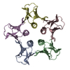
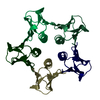
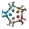
| Entry | Database: PDB / ID: 1c48 | ||||||
|---|---|---|---|---|---|---|---|
| Title | MUTATED SHIGA-LIKE TOXIN B SUBUNIT (G62T) | ||||||
 Components Components | PROTEIN (SHIGA-LIKE TOXIN I B SUBUNIT) | ||||||
 Keywords Keywords | TOXIN / RECEPTOR BINDING / PROTEIN-CARBOHYDRATE RECOGNITION / OB-FOLD | ||||||
| Function / homology |  Function and homology information Function and homology informationsymbiont-mediated modulation of host virulence / symbiont-mediated hemolysis of host erythrocyte / toxin activity / extracellular region Similarity search - Function | ||||||
| Biological species |  Phage h30 (virus) Phage h30 (virus) | ||||||
| Method |  X-RAY DIFFRACTION / SYNCHROTRON / MOLECULAR REPLACEMENT / Resolution: 1.6 Å X-RAY DIFFRACTION / SYNCHROTRON / MOLECULAR REPLACEMENT / Resolution: 1.6 Å | ||||||
 Authors Authors | Ling, H. / Bast, D. / Brunton, J.L. / Read, R.J. | ||||||
 Citation Citation | Journal: To be Published Title: Identification of the Primary Receptor Binding Site of Shiga-Like Toxin B Subunits: Structures of Mutated Shiga-Like Toxin I B-Pentamer with and without Bound Carbohydrate Authors: Ling, H. / Bast, D. / Brunton, J.L. / Read, R.J. #1: Journal: Biochemistry / Year: 1998Title: Structure of the Shiga-Like Toxin I B-Pentamer Complexed with an Analogue of its Receptor Gb3 Authors: Ling, H. / Boodhoo, A. / Hazes, B. / Cummings, M.D. / Armstrong, G.D. / Brunton, J.L. / Read, R.J. | ||||||
| History |
|
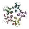
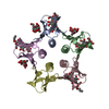
- Structure visualization
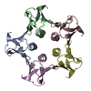
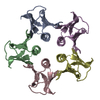
| Structure viewer | Molecule: MolmilJmol/JSmol |
|---|
- Downloads & links
-Download
| PDBx/mmCIF format | 1c48.cif.gz | 87.9 KB | Display | PDBx/mmCIF format |
|---|---|---|---|---|
| PDB format | pdb1c48.ent.gz | 67.1 KB | Display | PDB format |
| PDBx/mmJSON format | 1c48.json.gz | Tree view | PDBx/mmJSON format | |
| Others |  Other downloads Other downloads |
-Validation report
| Arichive directory | https://data.pdbj.org/pub/pdb/validation_reports/c4/1c48ftp://data.pdbj.org/pub/pdb/validation_reports/c4/1c48 | HTTPS FTP |
|---|
-Related structure data
| Related structure data | 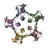 1c4qC  1bosS S: Starting model for refinement C: citing same article ( |
|---|---|
| Similar structure data |

-Links
 PDBj
PDBj
- Assembly
| Deposited unit | 
| ||||||||||||||||||||
|---|---|---|---|---|---|---|---|---|---|---|---|---|---|---|---|---|---|---|---|---|---|
| 1 |
| ||||||||||||||||||||
| Unit cell |
| ||||||||||||||||||||
| Noncrystallographic symmetry (NCS) | NCS oper:
|
-Components
| #1: Protein | Mass: 7742.686 Da / Num. of mol.: 5 / Fragment: RECEPTOR-BINDING DOMAIN / Mutation: G62T Source method: isolated from a genetically manipulated source Source: (gene. exp.) Phage h30 (virus) / Production host:  #2: Water | ChemComp-HOH / |  Mass: 18.015 Da / Num. of mol.: 348 / Source method: isolated from a natural source / Formula: H2O Mass: 18.015 Da / Num. of mol.: 348 / Source method: isolated from a natural source / Formula: H2OHas protein modification | Y | |
|---|
-Experimental details
-Experiment
| Experiment | Method: X-RAY DIFFRACTION / Number of used crystals: 1 |
|---|
- Sample preparation
| Crystal | Density Matthews: 2.07 Å3/Da / Density % sol: 40.56 % |
|---|---|
| Crystal grow | pH: 8.5 / Details: pH 8.5 |
-Data collection
| Diffraction | Mean temperature: 100 K |
|---|---|
| Diffraction source | Source: SYNCHROTRON / Site: NSLS  / Beamline: X12B / Wavelength: 0.978 / Beamline: X12B / Wavelength: 0.978 |
| Detector | Detector: CCD / Date: Mar 1, 1998 |
| Radiation | Protocol: SINGLE WAVELENGTH / Monochromatic (M) / Laue (L): M / Scattering type: x-ray |
| Radiation wavelength | Wavelength: 0.978 Å / Relative weight: 1 |
| Reflection | Resolution: 1.6→55 Å / Num. obs: 41266 / % possible obs: 98.8 % / Observed criterion σ(I): 0 / Redundancy: 4 % / Biso Wilson estimate: 21 Å2 / Rmerge(I) obs: 0.037 / Rsym value: 0.037 / Net I/σ(I): 3.7 |
| Reflection shell | Resolution: 1.6→1.65 Å / Rmerge(I) obs: 0.305 / Mean I/σ(I) obs: 2.1 / Rsym value: 0.305 / % possible all: 98.7 |
- Processing
| Software |
| ||||||||||||||||||||||||||||||||||||||||||||||||||||||||||||||||||||||||||||||||
|---|---|---|---|---|---|---|---|---|---|---|---|---|---|---|---|---|---|---|---|---|---|---|---|---|---|---|---|---|---|---|---|---|---|---|---|---|---|---|---|---|---|---|---|---|---|---|---|---|---|---|---|---|---|---|---|---|---|---|---|---|---|---|---|---|---|---|---|---|---|---|---|---|---|---|---|---|---|---|---|---|---|
| Refinement | Method to determine structure: MOLECULAR REPLACEMENT Starting model: 1BOS Resolution: 1.6→55 Å / Isotropic thermal model: RESTRAINED / Cross valid method: THROUGHOUT / σ(F): 0
| ||||||||||||||||||||||||||||||||||||||||||||||||||||||||||||||||||||||||||||||||
| Solvent computation | Bsol: 47.07 Å2 / ksol: 0.355 e/Å3 | ||||||||||||||||||||||||||||||||||||||||||||||||||||||||||||||||||||||||||||||||
| Displacement parameters | Biso mean: 20 Å2
| ||||||||||||||||||||||||||||||||||||||||||||||||||||||||||||||||||||||||||||||||
| Refinement step | Cycle: LAST / Resolution: 1.6→55 Å
| ||||||||||||||||||||||||||||||||||||||||||||||||||||||||||||||||||||||||||||||||
| Refine LS restraints |
| ||||||||||||||||||||||||||||||||||||||||||||||||||||||||||||||||||||||||||||||||
| Refine LS restraints NCS | NCS model details: RESTRAINTS | ||||||||||||||||||||||||||||||||||||||||||||||||||||||||||||||||||||||||||||||||
| LS refinement shell | Resolution: 1.6→1.65 Å / Total num. of bins used: 23
| ||||||||||||||||||||||||||||||||||||||||||||||||||||||||||||||||||||||||||||||||
| Xplor file |
|
