 Movie
Movie Controller

[English] 日本語
 Yorodumi
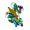
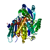
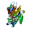
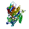
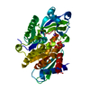
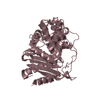
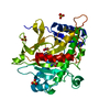
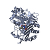
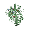
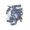
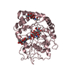
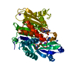
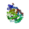
- PDB-2a9y: Crystal structure of T. gondii adenosine kinase complexed with N6... -
+ Open data

- Basic information
| Entry | Database: PDB / ID: 2a9y | ||||||
|---|---|---|---|---|---|---|---|
| Title | Crystal structure of T. gondii adenosine kinase complexed with N6-dimethyladenosine | ||||||
 Components Components | adenosine kinase | ||||||
 Keywords Keywords | SIGNALING PROTEIN / TRANSFERASE / ribokinase fold / alpha/beta | ||||||
| Function / homology |  Function and homology information Function and homology informationadenosine kinase / adenosine kinase activity / AMP salvage / purine ribonucleoside salvage / purine nucleobase metabolic process / phosphorylation / ATP binding / nucleus / metal ion binding / cytosol Similarity search - Function | ||||||
| Biological species |  | ||||||
| Method |  X-RAY DIFFRACTION / SYNCHROTRON / MOLECULAR REPLACEMENT / Resolution: 1.35 Å X-RAY DIFFRACTION / SYNCHROTRON / MOLECULAR REPLACEMENT / Resolution: 1.35 Å | ||||||
 Authors Authors | Zhang, Y. / el Kouni, M.H. / Ealick, S.E. | ||||||
 Citation Citation | Journal: Acta Crystallogr.,Sect.D / Year: 2007 Title: Substrate analogs induce an intermediate conformational change in Toxoplasma gondii adenosine kinase Authors: Zhang, Y. / El Kouni, M.H. / Ealick, S.E. | ||||||
| History |
|
- Structure visualization
| Structure viewer | Molecule: MolmilJmol/JSmol |
|---|
- Downloads & links
-Download
| PDBx/mmCIF format | 2a9y.cif.gz | 174.7 KB | Display | PDBx/mmCIF format |
|---|---|---|---|---|
| PDB format | pdb2a9y.ent.gz | 135.1 KB | Display | PDB format |
| PDBx/mmJSON format | 2a9y.json.gz | Tree view | PDBx/mmJSON format | |
| Others |  Other downloads Other downloads |
-Validation report
| Summary document | 2a9y_validation.pdf.gz | 1007.7 KB | Display | wwPDB validaton report |
|---|---|---|---|---|
| Full document | 2a9y_full_validation.pdf.gz | 1013.5 KB | Display | |
| Data in XML | 2a9y_validation.xml.gz | 19.9 KB | Display | |
| Data in CIF | 2a9y_validation.cif.gz | 30.7 KB | Display | |
| Arichive directory | https://data.pdbj.org/pub/pdb/validation_reports/a9/2a9yftp://data.pdbj.org/pub/pdb/validation_reports/a9/2a9y | HTTPS FTP |
-Related structure data
| Related structure data | 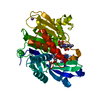 2a9zC 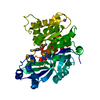 2aa0C  2ab8C 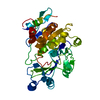 1lioS S: Starting model for refinement C: citing same article ( |
|---|---|
| Similar structure data |


-Links
 PDBj
PDBj- Assembly
| Deposited unit | 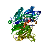
| ||||||||||
|---|---|---|---|---|---|---|---|---|---|---|---|
| 1 |
| ||||||||||
| Unit cell |
|
-Components
-Protein , 1 types, 1 molecules A
| #1: Protein | Mass: 40584.105 Da / Num. of mol.: 1 / Mutation: G270S, S360F, L361T, P362S, C363G Source method: isolated from a genetically manipulated source Source: (gene. exp.)  |
|---|
-Non-polymers , 5 types, 410 molecules 


| #2: Chemical | ChemComp-CL /  Mass: 35.453 Da / Num. of mol.: 1 / Source method: obtained synthetically / Formula: Cl Mass: 35.453 Da / Num. of mol.: 1 / Source method: obtained synthetically / Formula: Cl | ||||||
|---|---|---|---|---|---|---|---|
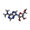
| #3: Chemical |  Mass: 22.990 Da / Num. of mol.: 2 / Source method: obtained synthetically / Formula: Na Mass: 22.990 Da / Num. of mol.: 2 / Source method: obtained synthetically / Formula: Na#4: Chemical | ChemComp-ACT / |  Mass: 59.044 Da / Num. of mol.: 1 / Source method: obtained synthetically / Formula: C2H3O2 Mass: 59.044 Da / Num. of mol.: 1 / Source method: obtained synthetically / Formula: C2H3O2#5: Chemical |  Mass: 295.294 Da / Num. of mol.: 2 / Source method: obtained synthetically / Formula: C12H17N5O4 Mass: 295.294 Da / Num. of mol.: 2 / Source method: obtained synthetically / Formula: C12H17N5O4#6: Water | ChemComp-HOH / | Mass: 18.015 Da / Num. of mol.: 404 / Source method: isolated from a natural source / Formula: H2O |
-Experimental details
-Experiment
| Experiment | Method: X-RAY DIFFRACTION / Number of used crystals: 1 |
|---|
- Sample preparation
| Crystal | Density Matthews: 2.1 Å3/Da / Density % sol: 41.8 % |
|---|---|
| Crystal grow | Temperature: 295 K / Method: vapor diffusion, hanging drop / pH: 6.5 Details: PEG 4000, ammonium acetate, sodium citrate, pH 6.5, VAPOR DIFFUSION, HANGING DROP, temperature 295K |
-Data collection
| Diffraction | Mean temperature: 100 K |
|---|---|
| Diffraction source | Source: SYNCHROTRON / Site: CHESS  / Beamline: F2 / Wavelength: 0.9795 Å / Beamline: F2 / Wavelength: 0.9795 Å |
| Detector | Type: ADSC QUANTUM 210 / Detector: CCD / Date: Nov 29, 2004 |
| Radiation | Monochromator: Si(111) crystals / Protocol: SINGLE WAVELENGTH / Monochromatic (M) / Laue (L): M / Scattering type: x-ray |
| Radiation wavelength | Wavelength: 0.9795 Å / Relative weight: 1 |
| Reflection | Resolution: 1.35→50 Å / Num. all: 72440 / Num. obs: 72440 / % possible obs: 95.5 % / Observed criterion σ(F): 0 / Observed criterion σ(I): 0 / Redundancy: 4.6 % / Rsym value: 0.06 / Net I/σ(I): 22.7 |
| Reflection shell | Resolution: 1.35→1.4 Å / Redundancy: 2.9 % / Mean I/σ(I) obs: 2.1 / Num. unique all: 5480 / Rsym value: 0.378 / % possible all: 73.2 |
- Processing
| Software |
| |||||||||||||||||||||||||||||||||
|---|---|---|---|---|---|---|---|---|---|---|---|---|---|---|---|---|---|---|---|---|---|---|---|---|---|---|---|---|---|---|---|---|---|---|
| Refinement | Method to determine structure: MOLECULAR REPLACEMENT Starting model: PDB entry 1LIO Resolution: 1.35→10 Å / Num. parameters: 28776 / Num. restraintsaints: 36552 / Cross valid method: FREE R / σ(F): 0 / Stereochemistry target values: Engh & Huber Details: ANISOTROPIC REFINEMENT REDUCED FREE R (NO CUTOFF) BY 4.1%
| |||||||||||||||||||||||||||||||||
| Refine analyze | Num. disordered residues: 30 / Occupancy sum hydrogen: 0 / Occupancy sum non hydrogen: 3046 | |||||||||||||||||||||||||||||||||
| Refinement step | Cycle: LAST / Resolution: 1.35→10 Å
| |||||||||||||||||||||||||||||||||
| Refine LS restraints |
| |||||||||||||||||||||||||||||||||
| LS refinement shell | Resolution: 1.35→1.42 Å /
|
