 ムービー
ムービー コントローラー


+ データを開く

- 基本情報
基本情報
| 登録情報 | データベース: EMDB / ID: EMD-2913 | |||||||||
|---|---|---|---|---|---|---|---|---|---|---|
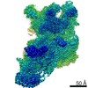
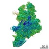
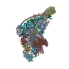
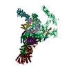
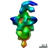
| タイトル | Cryo-EM reconstruction of the mammalian 28S mitoribosomal subunit | |||||||||
 マップデータ マップデータ | Reconstruction of the porcine 28S mitoribosomal subunit | |||||||||
 試料 試料 |
| |||||||||
 キーワード キーワード | mammalian mitochondrial ribosome / 28S small ribosomal subunit / translation / ribosomal proteins / rRNA / tRNA / mRNA / decoding | |||||||||
| 機能・相同性 |  機能・相同性情報 機能・相同性情報Mitochondrial translation elongation / Mitochondrial translation termination / mitochondrial ribosome assembly / Mitochondrial protein degradation / mitochondrial ribosome / mitochondrial small ribosomal subunit / mitochondrial translation / ribosomal small subunit binding / cell junction / regulation of translation ...Mitochondrial translation elongation / Mitochondrial translation termination / mitochondrial ribosome assembly / Mitochondrial protein degradation / mitochondrial ribosome / mitochondrial small ribosomal subunit / mitochondrial translation / ribosomal small subunit binding / cell junction / regulation of translation / small ribosomal subunit / small ribosomal subunit rRNA binding / rRNA binding / ribosome / structural constituent of ribosome / translation / ribonucleoprotein complex / apoptotic process / mitochondrion / RNA binding / nucleoplasm / cytoplasm 類似検索 - 分子機能 | |||||||||
| 生物種 |  | |||||||||
| 手法 | 単粒子再構成法 / クライオ電子顕微鏡法 / 解像度: 3.6 Å | |||||||||
 データ登録者 データ登録者 | Greber BJ / Bieri P / Leibundgut M / Leitner A / Aebersold R / Boehringer D / Ban N | |||||||||
 引用 引用 | ジャーナル: Science / 年: 2015 タイトル: Ribosome. The complete structure of the 55S mammalian mitochondrial ribosome. 著者: Basil J Greber / Philipp Bieri / Marc Leibundgut / Alexander Leitner / Ruedi Aebersold / Daniel Boehringer / Nenad Ban /  要旨: Mammalian mitochondrial ribosomes (mitoribosomes) synthesize mitochondrially encoded membrane proteins that are critical for mitochondrial function. Here we present the complete atomic structure of ...Mammalian mitochondrial ribosomes (mitoribosomes) synthesize mitochondrially encoded membrane proteins that are critical for mitochondrial function. Here we present the complete atomic structure of the porcine 55S mitoribosome at 3.8 angstrom resolution by cryo-electron microscopy and chemical cross-linking/mass spectrometry. The structure of the 28S subunit in the complex was resolved at 3.6 angstrom resolution by focused alignment, which allowed building of a detailed atomic structure including all of its 15 mitoribosomal-specific proteins. The structure reveals the intersubunit contacts in the 55S mitoribosome, the molecular architecture of the mitoribosomal messenger RNA (mRNA) binding channel and its interaction with transfer RNAs, and provides insight into the highly specialized mechanism of mRNA recruitment to the 28S subunit. Furthermore, the structure contributes to a mechanistic understanding of aminoglycoside ototoxicity. | |||||||||
| 履歴 |
|
- 構造の表示
構造の表示
| ムービー |
ムービービューア |
|---|---|
| 構造ビューア | EMマップ: SurfViewMolmilJmol/JSmol |
| 添付画像 |

- ダウンロードとリンク
-EMDBアーカイブ
| マップデータ | emd_2913.map.gz | 7.4 MB | EMDBマップデータ形式 | |
|---|---|---|---|---|
| ヘッダ (付随情報) | emd-2913-v30.xmlemd-2913.xml | 11.3 KB 11.3 KB | 表示 表示 | EMDBヘッダ |
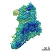
| 画像 | 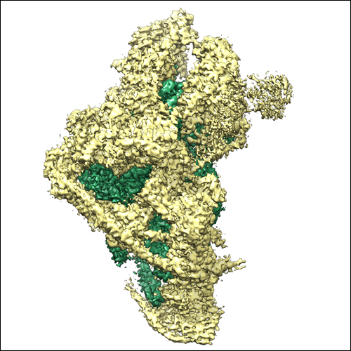 EMDB_2913_28S_500px.png EMDB_2913_28S_500px.png | 233 KB | ||
| アーカイブディレクトリ |  http://ftp.pdbj.org/pub/emdb/structures/EMD-2913ftp://ftp.pdbj.org/pub/emdb/structures/EMD-2913 http://ftp.pdbj.org/pub/emdb/structures/EMD-2913ftp://ftp.pdbj.org/pub/emdb/structures/EMD-2913 | HTTPS FTP |
-検証レポート
| 文書・要旨 | emd_2913_validation.pdf.gz | 248.5 KB | 表示 | EMDB検証レポート |
|---|---|---|---|---|
| 文書・詳細版 | emd_2913_full_validation.pdf.gz | 247.6 KB | 表示 | |
| XML形式データ | emd_2913_validation.xml.gz | 5.8 KB | 表示 | |
| アーカイブディレクトリ | https://ftp.pdbj.org/pub/emdb/validation_reports/EMD-2913ftp://ftp.pdbj.org/pub/emdb/validation_reports/EMD-2913 | HTTPS FTP |
-関連構造データ
-リンク
| EMDBのページ | EMDB (EBI/PDBe) / EMDataResource |
|---|---|
| 「今月の分子」の関連する項目 |


-マップ
| ファイル | ダウンロード / ファイル: emd_2913.map.gz / 形式: CCP4 / 大きさ: 37.5 MB / タイプ: IMAGE STORED AS FLOATING POINT NUMBER (4 BYTES) | ||||||||||||||||||||||||||||||||||||||||||||||||||||||||||||||||||||
|---|---|---|---|---|---|---|---|---|---|---|---|---|---|---|---|---|---|---|---|---|---|---|---|---|---|---|---|---|---|---|---|---|---|---|---|---|---|---|---|---|---|---|---|---|---|---|---|---|---|---|---|---|---|---|---|---|---|---|---|---|---|---|---|---|---|---|---|---|---|
| 注釈 | Reconstruction of the porcine 28S mitoribosomal subunit | ||||||||||||||||||||||||||||||||||||||||||||||||||||||||||||||||||||
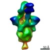
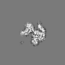
| 投影像・断面図 | 画像のコントロール
画像は Spider により作成 | ||||||||||||||||||||||||||||||||||||||||||||||||||||||||||||||||||||
| ボクセルのサイズ | X=Y=Z: 1.39 Å | ||||||||||||||||||||||||||||||||||||||||||||||||||||||||||||||||||||
| 密度 |
| ||||||||||||||||||||||||||||||||||||||||||||||||||||||||||||||||||||
| 対称性 | 空間群: 1 | ||||||||||||||||||||||||||||||||||||||||||||||||||||||||||||||||||||
| 詳細 | EMDB XML:
CCP4マップ ヘッダ情報:
| ||||||||||||||||||||||||||||||||||||||||||||||||||||||||||||||||||||
 Z (Sec.)
 Y (Row.)
 X (Col.)


-添付データ
- 試料の構成要素
-全体 : 28S mammalian mitochondrial ribosome
| 全体 | 名称: 28S mammalian mitochondrial ribosome |
|---|---|
| 要素 |
|
-超分子 #1000: 28S mammalian mitochondrial ribosome
| 超分子 | 名称: 28S mammalian mitochondrial ribosome / タイプ: sample / ID: 1000 / 集合状態: monomer / Number unique components: 1 |
|---|---|
| 分子量 | 理論値: 2.7 MDa |
-超分子 #1: 28S mammalian mitochondrial ribosome subunit
| 超分子 | 名称: 28S mammalian mitochondrial ribosome subunit / タイプ: complex / ID: 1 / 組換発現: No Ribosome-details: ribosome-eukaryote: SSU mitochondrial 28S, SSU mitochondrial RNA 12S |
|---|---|
| Ref GO | 0: GO:0005761 |
| 由来(天然) | 生物種: |
| 分子量 | 理論値: 1.1 MDa |
-実験情報
-構造解析
| 手法 | クライオ電子顕微鏡法 |
|---|---|
 解析 解析 | 単粒子再構成法 |
| 試料の集合状態 | particle |
-試料調製
| 緩衝液 | pH: 7.4 / 詳細: 20 mM HEPES-KOH, 50 mM KCl, 1 mM DTT, 40 mM MgCl2 |
|---|---|
| グリッド | 詳細: 200 mesh Quantifoil R 2/2 holey carbon grids with a thin continuous carbon support film applied |
| 凍結 | 凍結剤: ETHANE-PROPANE MIXTURE / 装置: HOMEMADE PLUNGER |
- 電子顕微鏡法 #1
| Microscopy ID | 1 |
|---|---|
| 顕微鏡 | FEI TITAN KRIOS |
| 詳細 | movie mode readout in FEI EPU: 7 frames per exposure |
| 日付 | 2014年5月3日 |
| 撮影 | カテゴリ: CCD フィルム・検出器のモデル: FEI FALCON II (4k x 4k) デジタル化 - サンプリング間隔: 14 µm / 平均電子線量: 20 e/Å2 詳細: movie mode readout in FEI EPU: 7 frames per exposure |
| 電子線 | 加速電圧: 300 kV / 電子線源:  FIELD EMISSION GUN FIELD EMISSION GUN |
| 電子光学系 | 倍率(補正後): 100720 / 照射モード: FLOOD BEAM / 撮影モード: BRIGHT FIELD / Cs: 2.7 mm / 最大 デフォーカス(公称値): 3.0 µm / 最小 デフォーカス(公称値): 0.8 µm / 倍率(公称値): 59000 |
| 試料ステージ | 試料ホルダーモデル: FEI TITAN KRIOS AUTOGRID HOLDER |
| 実験機器 |  モデル: Titan Krios / 画像提供: FEI Company |
-電子顕微鏡法 #2
| Microscopy ID | 2 |
|---|---|
| 顕微鏡 | FEI TITAN KRIOS |
| 詳細 | movie mode readout in FEI EPU: 7 frames per exposure |
| 日付 | 2014年5月30日 |
| 撮影 | カテゴリ: CCD フィルム・検出器のモデル: FEI FALCON II (4k x 4k) デジタル化 - サンプリング間隔: 14 µm / 平均電子線量: 20 e/Å2 詳細: movie mode readout in FEI EPU: 7 frames per exposure |
| 電子線 | 加速電圧: 300 kV / 電子線源: FIELD EMISSION GUN |
| 電子光学系 | 倍率(補正後): 100720 / 照射モード: FLOOD BEAM / 撮影モード: BRIGHT FIELD / Cs: 2.7 mm / 最大 デフォーカス(公称値): 3.0 µm / 最小 デフォーカス(公称値): 0.8 µm / 倍率(公称値): 59000 |
| 試料ステージ | 試料ホルダーモデル: FEI TITAN KRIOS AUTOGRID HOLDER |
| 実験機器 | モデル: Titan Krios / 画像提供: FEI Company |
-画像解析
| 詳細 | Particles were selected in batchboxer (EMAN 1.9) and extracted using RELION 1.2. |
|---|---|
| CTF補正 | 詳細: per detector frame |
| 最終 再構成 | 想定した対称性 - 点群: C1 (非対称) / アルゴリズム: OTHER / 解像度のタイプ: BY AUTHOR / 解像度: 3.6 Å / 解像度の算出法: OTHER / ソフトウェア - 名称: CTFFIND3, RELION, 1.2, 1.3 / 使用した粒子像数: 78783 |
-原子モデル構築 1
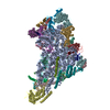
| 初期モデル | PDB ID:  3v2c |
|---|---|
| ソフトウェア | 名称: Chimera |
| 詳細 | The initial coordinate model was fitted into the cryo-EM density using Chimera. The model was then extended and completely rebuilt using COOT (RNA) and O (proteins) aided by PHYRE models for ribosomal proteins. |
| 精密化 | 空間: REAL / プロトコル: RIGID BODY FIT |
| 得られたモデル | 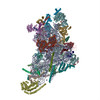 PDB-5aj3: |
