National Institutes of Health/National Heart, Lung, and Blood Institute (NIH/NHLBI)
R01HL036153
米国
引用
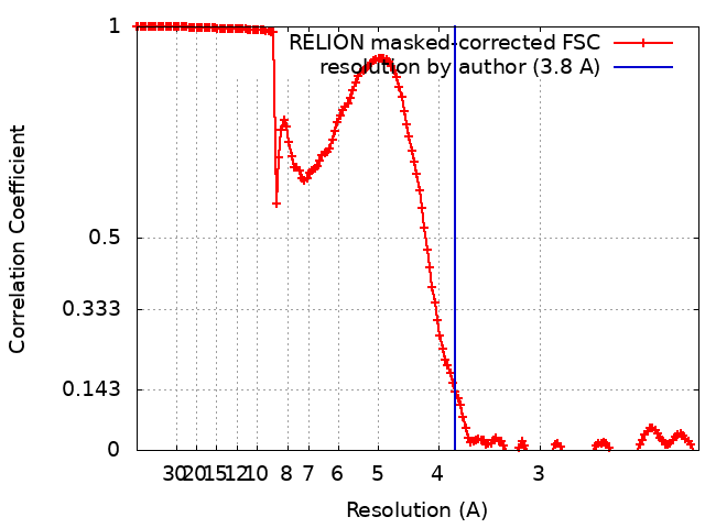
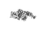
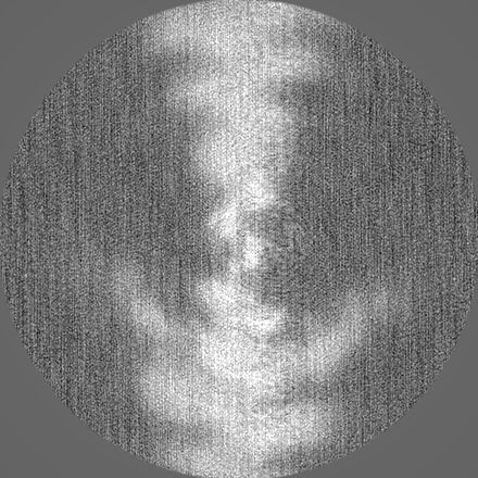
ジャーナル: J Gen Physiol / 年: 2023 タイトル: Conformational changes linked to ADP release from human cardiac myosin bound to actin-tropomyosin. 著者: Matthew H Doran / Michael J Rynkiewicz / David Rasicci / Skylar M L Bodt / Meaghan E Barry / Esther Bullitt / Christopher M Yengo / Jeffrey R Moore / William Lehman / 要旨: Following binding to the thin filament, β-cardiac myosin couples ATP-hydrolysis to conformational rearrangements in the myosin motor that drive myofilament sliding and cardiac ventricular ...Following binding to the thin filament, β-cardiac myosin couples ATP-hydrolysis to conformational rearrangements in the myosin motor that drive myofilament sliding and cardiac ventricular contraction. However, key features of the cardiac-specific actin-myosin interaction remain uncertain, including the structural effect of ADP release from myosin, which is rate-limiting during force generation. In fact, ADP release slows under experimental load or in the intact heart due to the afterload, thereby adjusting cardiac muscle power output to meet physiological demands. To further elucidate the structural basis of this fundamental process, we used a combination of cryo-EM reconstruction methodologies to determine structures of the human cardiac actin-myosin-tropomyosin filament complex at better than 3.4 Å-resolution in the presence and in the absence of Mg2+·ADP. Focused refinements of the myosin motor head and its essential light chains in these reconstructions reveal that small changes in the nucleotide-binding site are coupled to significant rigid body movements of the myosin converter domain and a 16-degree lever arm swing. Our structures provide a mechanistic framework to understand the effect of ADP binding and release on human cardiac β-myosin, and offer insights into the force-sensing mechanism displayed by the cardiac myosin motor.
超分子 #1: Complex of the human beta cardiac myosin II in the rigor form wit...
超分子
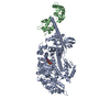
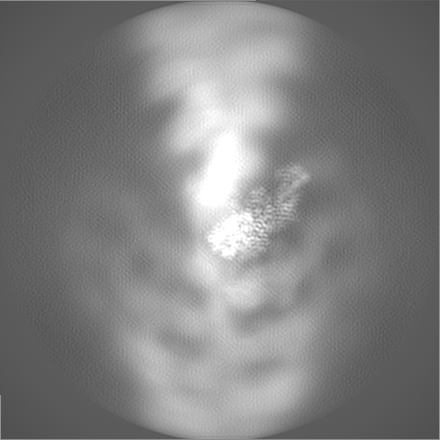
名称: Complex of the human beta cardiac myosin II in the rigor form with the associated essential light chain. タイプ: complex / ID: 1 / 親要素: 0 / 含まれる分子: all
-
超分子 #2: Human beta-cardiac myosin II
超分子
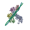
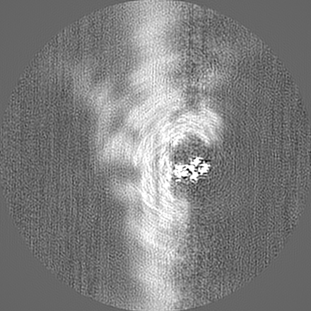
名称: Human beta-cardiac myosin II / タイプ: complex / ID: 2 / 親要素: 1 / 含まれる分子: #1
 ムービー
ムービー コントローラー


 データを開く
 基本情報
基本情報
 マップデータ
 試料
試料 キーワード
キーワード 機能・相同性情報
 Homo sapiens (ヒト) /
 データ登録者
 米国, 1件
  引用
引用 構造の表示
構造の表示
 ダウンロードとリンク
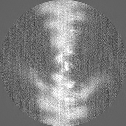
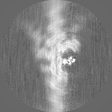
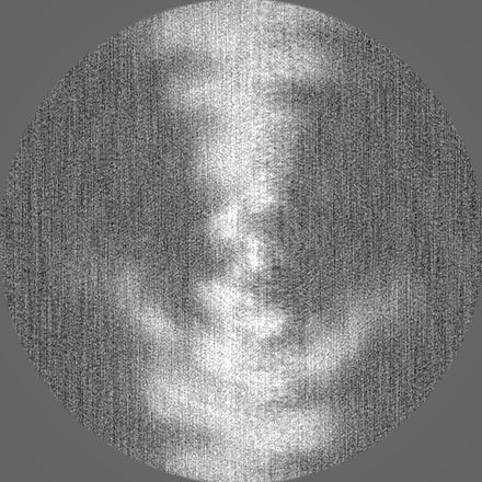
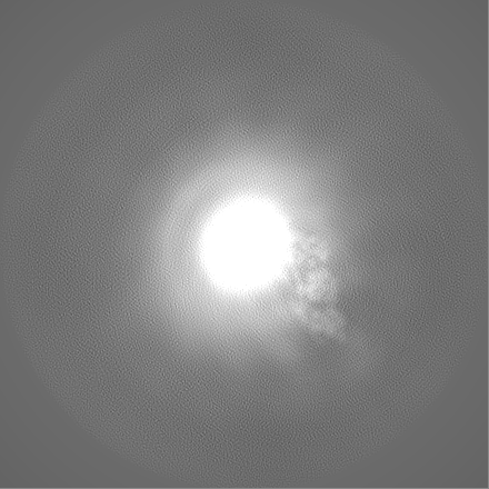
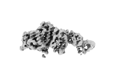 emd_28080.png
 http://ftp.pdbj.org/pub/emdb/structures/EMD-28080
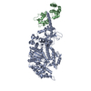


 Z
Z Y
Y X
X


 試料の構成要素
 解析
解析 電子顕微鏡法
 FIELD EMISSION GUN