 Movie
Movie Controller

[English] 日本語
 Yorodumi
- EMDB-25809: Sub-tomogram averaged map of HIV-Env with Gag layer from immature VLPs -
+ Open data

- Basic information
| Entry | Database: EMDB / ID: EMD-25809 | ||||||||||||
|---|---|---|---|---|---|---|---|---|---|---|---|---|---|
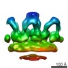
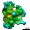
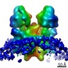
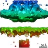
| Title | Sub-tomogram averaged map of HIV-Env with Gag layer from immature VLPs | ||||||||||||
 Map data Map data | Immature hVLP Env (relaxed C1 sym) with Gag layer - half map | ||||||||||||
 Sample Sample |
| ||||||||||||
 Keywords Keywords | HIV-1 / envelope protein / glycoprotein / Env / membrane bound / VIRAL PROTEIN / Gag | ||||||||||||
| Function / homology |  Function and homology information Function and homology informationvirus-mediated perturbation of host defense response => GO:0019049 / : / stimulatory C-type lectin receptor signaling pathway / positive regulation of plasma membrane raft polarization / positive regulation of receptor clustering / positive regulation of establishment of T cell polarity / host cell endosome membrane / clathrin-dependent endocytosis of virus by host cell / membrane => GO:0016020 / viral protein processing ...virus-mediated perturbation of host defense response => GO:0019049 / : / stimulatory C-type lectin receptor signaling pathway / positive regulation of plasma membrane raft polarization / positive regulation of receptor clustering / positive regulation of establishment of T cell polarity / host cell endosome membrane / clathrin-dependent endocytosis of virus by host cell / membrane => GO:0016020 / viral protein processing / fusion of virus membrane with host plasma membrane / fusion of virus membrane with host endosome membrane / viral envelope / virion attachment to host cell / host cell plasma membrane / structural molecule activity / virion membrane / plasma membrane Similarity search - Function | ||||||||||||
| Biological species |   Human immunodeficiency virus 1 Human immunodeficiency virus 1 | ||||||||||||
| Method | subtomogram averaging / cryo EM / Resolution: 34.0 Å | ||||||||||||
 Authors Authors | Mangala Prasad V / Lee KK | ||||||||||||
| Funding support |  United States, 3 items United States, 3 items
| ||||||||||||
 Citation Citation | Journal: Cell / Year: 2022 Title: Cryo-ET of Env on intact HIV virions reveals structural variation and positioning on the Gag lattice. Authors: Vidya Mangala Prasad / Daniel P Leaman / Klaus N Lovendahl / Jacob T Croft / Mark A Benhaim / Edgar A Hodge / Michael B Zwick / Kelly K Lee / Abstract: HIV-1 Env mediates viral entry into host cells and is the sole target for neutralizing antibodies. However, Env structure and organization in its native virion context has eluded detailed ...HIV-1 Env mediates viral entry into host cells and is the sole target for neutralizing antibodies. However, Env structure and organization in its native virion context has eluded detailed characterization. Here, we used cryo-electron tomography to analyze Env in mature and immature HIV-1 particles. Immature particles showed distinct Env positioning relative to the underlying Gag lattice, providing insights into long-standing questions about Env incorporation. A 9.1-Å sub-tomogram-averaged reconstruction of virion-bound Env in conjunction with structural mass spectrometry revealed unexpected features, including a variable central core of the gp41 subunit, heterogeneous glycosylation between protomers, and a flexible stalk that allows Env tilting and variable exposure of neutralizing epitopes. Together, our results provide an integrative understanding of HIV assembly and structural variation in Env antigen presentation. | ||||||||||||
| History |
|
- Structure visualization
| Movie |
Movie viewer |
|---|---|
| Structure viewer | EM map: SurfViewMolmilJmol/JSmol |
| Supplemental images |

- Downloads & links
-EMDB archive
| Map data | emd_25809.map.gz | 118.9 KB | EMDB map data format | |
|---|---|---|---|---|
| Header (meta data) | emd-25809-v30.xmlemd-25809.xml | 16.2 KB 16.2 KB | Display Display | EMDB header |
| FSC (resolution estimation) | emd_25809_fsc.xml | 1.1 KB | Display | FSC data file |
| Images |  emd_25809.png emd_25809.png | 74.5 KB | ||
| Filedesc metadata | emd-25809.cif.gz | 5.1 KB | ||
| Others | emd_25809_additional_1.map.gz | 118.9 KB | ||
| Archive directory |  http://ftp.pdbj.org/pub/emdb/structures/EMD-25809ftp://ftp.pdbj.org/pub/emdb/structures/EMD-25809 http://ftp.pdbj.org/pub/emdb/structures/EMD-25809ftp://ftp.pdbj.org/pub/emdb/structures/EMD-25809 | HTTPS FTP |
-Validation report
| Summary document | emd_25809_validation.pdf.gz | 495.1 KB | Display | EMDB validaton report |
|---|---|---|---|---|
| Full document | emd_25809_full_validation.pdf.gz | 494.7 KB | Display | |
| Data in XML | emd_25809_validation.xml.gz | 5.8 KB | Display | |
| Data in CIF | emd_25809_validation.cif.gz | 6.7 KB | Display | |
| Arichive directory | https://ftp.pdbj.org/pub/emdb/validation_reports/EMD-25809ftp://ftp.pdbj.org/pub/emdb/validation_reports/EMD-25809 | HTTPS FTP |
-Related structure data


-Links
| EMDB pages | EMDB (EBI/PDBe) / EMDataResource |
|---|---|
| Related items in Molecule of the Month |


-Map
| File | Download / File: emd_25809.map.gz / Format: CCP4 / Size: 128.9 KB / Type: IMAGE STORED AS FLOATING POINT NUMBER (4 BYTES) | ||||||||||||||||||||||||||||||||||||||||||||||||||||||||||||||||||||
|---|---|---|---|---|---|---|---|---|---|---|---|---|---|---|---|---|---|---|---|---|---|---|---|---|---|---|---|---|---|---|---|---|---|---|---|---|---|---|---|---|---|---|---|---|---|---|---|---|---|---|---|---|---|---|---|---|---|---|---|---|---|---|---|---|---|---|---|---|---|
| Annotation | Immature hVLP Env (relaxed C1 sym) with Gag layer - half map | ||||||||||||||||||||||||||||||||||||||||||||||||||||||||||||||||||||
| Voxel size | X=Y=Z: 10.32 Å | ||||||||||||||||||||||||||||||||||||||||||||||||||||||||||||||||||||
| Density |
| ||||||||||||||||||||||||||||||||||||||||||||||||||||||||||||||||||||
| Symmetry | Space group: 1 | ||||||||||||||||||||||||||||||||||||||||||||||||||||||||||||||||||||
| Details | EMDB XML:
CCP4 map header:
| ||||||||||||||||||||||||||||||||||||||||||||||||||||||||||||||||||||
-Supplemental data
-Additional map: Immature hVLP Env (relaxed C1 sym) with Gag layer - full combined map
| File | emd_25809_additional_1.map | ||||||||||||
|---|---|---|---|---|---|---|---|---|---|---|---|---|---|
| Annotation | Immature hVLP Env (relaxed C1 sym) with Gag layer - full combined map | ||||||||||||
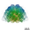
| Projections & Slices |
| ||||||||||||
| Density Histograms |
 Z
Z Y
Y X
X


- Sample components
-Entire : Human immunodeficiency virus 1
| Entire | Name: Human immunodeficiency virus 1 |
|---|---|
| Components |
|
-Supramolecule #1: Human immunodeficiency virus 1
| Supramolecule | Name: Human immunodeficiency virus 1 / type: virus / ID: 1 / Parent: 0 / Macromolecule list: #1 Details: HIV-1 ADA.CM Env glycoprotein displayed on high Env expressing VLPs NCBI-ID: 11676 / Sci species name: Human immunodeficiency virus 1 / Virus type: VIRUS-LIKE PARTICLE / Virus isolate: STRAIN / Virus enveloped: Yes / Virus empty: No |
|---|---|
| Host (natural) | Organism:  Homo sapiens (human) / Strain: ADA.CM Homo sapiens (human) / Strain: ADA.CM |
| Molecular weight | Theoretical: 400 KDa |
| Virus shell | Shell ID: 1 / Name: Outer membrane envelope / Diameter: 1000.0 Å |
-Experimental details
-Structure determination
| Method | cryo EM |
|---|---|
 Processing Processing | subtomogram averaging |
| Aggregation state | particle |
-Sample preparation
| Buffer | pH: 7.4 / Component - Concentration: 1.0 X / Component - Name: Phosphate buffer saline / Details: 1X PBS at pH 7.4 |
|---|---|
| Grid | Model: Quantifoil R1.2/1.3 / Material: COPPER / Mesh: 400 / Support film - Material: CARBON / Support film - topology: HOLEY / Pretreatment - Type: GLOW DISCHARGE / Pretreatment - Time: 30 sec. / Pretreatment - Atmosphere: OTHER |
| Vitrification | Cryogen name: ETHANE / Chamber humidity: 100 % / Chamber temperature: 277 K / Instrument: FEI VITROBOT MARK IV / Details: 3ul sample, blotting time of 4-5 seconds. |
| Details | Immature VLP particles displaying nearly full-length Env glycoprotein on membrane surface |
- Electron microscopy
| Microscope | FEI TITAN KRIOS |
|---|---|
| Image recording | Film or detector model: GATAN K2 SUMMIT (4k x 4k) / Detector mode: COUNTING / Average electron dose: 2.0 e/Å2 |
| Electron beam | Acceleration voltage: 300 kV / Electron source:  FIELD EMISSION GUN FIELD EMISSION GUN |
| Electron optics | Illumination mode: FLOOD BEAM / Imaging mode: BRIGHT FIELD / Cs: 2.7 mm / Nominal defocus max: 5.0 µm / Nominal defocus min: 2.5 µm / Nominal magnification: 58000 |
| Sample stage | Specimen holder model: FEI TITAN KRIOS AUTOGRID HOLDER / Cooling holder cryogen: NITROGEN |
| Experimental equipment |  Model: Titan Krios / Image courtesy: FEI Company |
+Image processing

-Atomic model buiding 1
| Initial model | PDB ID: Chain - Source name: PDB / Chain - Initial model type: experimental model |
|---|---|
| Details | hVLP-Env on membrane surface |
| Refinement | Space: REAL / Protocol: RIGID BODY FIT / Target criteria: Correlation coefficient |
-Atomic model buiding 2
| Initial model | PDB ID: Chain - Source name: PDB / Chain - Initial model type: experimental model |
|---|---|
| Details | Gag-CA hexamer structure for inner protein layer under membrane bilayer |
| Refinement | Protocol: RIGID BODY FIT / Target criteria: Correlation coefficient |

