 Movie
Movie Controller

[English] 日本語
 Yorodumi
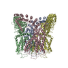
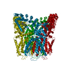
- EMDB-25178: Sub-tomogram averaged structure of HIV-1 Envelope protein in nati... -
+ Open data

- Basic information
| Entry | Database: EMDB / ID: EMD-25178 | ||||||||||||
|---|---|---|---|---|---|---|---|---|---|---|---|---|---|
| Title | Sub-tomogram averaged structure of HIV-1 Envelope protein in native membrane | ||||||||||||
 Map data Map data | |||||||||||||
 Sample Sample |
| ||||||||||||
 Keywords Keywords | HIV-1 / envelope protein / glycoprotein / Env / membrane bound / VIRAL PROTEIN | ||||||||||||
| Function / homology |  Function and homology information Function and homology informationsymbiont-mediated perturbation of host defense response / positive regulation of plasma membrane raft polarization / positive regulation of receptor clustering / host cell endosome membrane / clathrin-dependent endocytosis of virus by host cell / viral protein processing / fusion of virus membrane with host plasma membrane / fusion of virus membrane with host endosome membrane / viral envelope / virion attachment to host cell ...symbiont-mediated perturbation of host defense response / positive regulation of plasma membrane raft polarization / positive regulation of receptor clustering / host cell endosome membrane / clathrin-dependent endocytosis of virus by host cell / viral protein processing / fusion of virus membrane with host plasma membrane / fusion of virus membrane with host endosome membrane / viral envelope / virion attachment to host cell / host cell plasma membrane / virion membrane / structural molecule activity / membrane Similarity search - Function | ||||||||||||
| Biological species |   Human immunodeficiency virus 1 Human immunodeficiency virus 1 | ||||||||||||
| Method | subtomogram averaging / cryo EM / Resolution: 9.1 Å | ||||||||||||
 Authors Authors | Mangala Prasad V / Lee KK | ||||||||||||
| Funding support |  United States, 3 items United States, 3 items
| ||||||||||||
 Citation Citation | Journal: Cell / Year: 2022 Title: Cryo-ET of Env on intact HIV virions reveals structural variation and positioning on the Gag lattice. Authors: Vidya Mangala Prasad / Daniel P Leaman / Klaus N Lovendahl / Jacob T Croft / Mark A Benhaim / Edgar A Hodge / Michael B Zwick / Kelly K Lee / Abstract: HIV-1 Env mediates viral entry into host cells and is the sole target for neutralizing antibodies. However, Env structure and organization in its native virion context has eluded detailed ...HIV-1 Env mediates viral entry into host cells and is the sole target for neutralizing antibodies. However, Env structure and organization in its native virion context has eluded detailed characterization. Here, we used cryo-electron tomography to analyze Env in mature and immature HIV-1 particles. Immature particles showed distinct Env positioning relative to the underlying Gag lattice, providing insights into long-standing questions about Env incorporation. A 9.1-Å sub-tomogram-averaged reconstruction of virion-bound Env in conjunction with structural mass spectrometry revealed unexpected features, including a variable central core of the gp41 subunit, heterogeneous glycosylation between protomers, and a flexible stalk that allows Env tilting and variable exposure of neutralizing epitopes. Together, our results provide an integrative understanding of HIV assembly and structural variation in Env antigen presentation. | ||||||||||||
| History |
|
- Structure visualization
| Movie |
Movie viewer |
|---|---|
| Structure viewer | EM map: SurfViewMolmilJmol/JSmol |
| Supplemental images |

- Downloads & links
-EMDB archive
| Map data | emd_25178.map.gz | 855.6 KB | EMDB map data format | |
|---|---|---|---|---|
| Header (meta data) | emd-25178-v30.xmlemd-25178.xml | 17.8 KB 17.8 KB | Display Display | EMDB header |
| FSC (resolution estimation) | emd_25178_fsc.xml | 4.3 KB | Display | FSC data file |
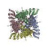
| Images | 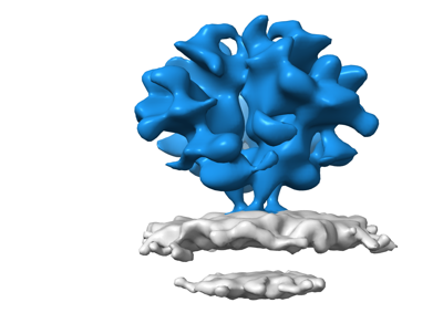 emd_25178.png emd_25178.png | 57.5 KB | ||
| Filedesc metadata | emd-25178.cif.gz | 7.4 KB | ||
| Archive directory |  http://ftp.pdbj.org/pub/emdb/structures/EMD-25178ftp://ftp.pdbj.org/pub/emdb/structures/EMD-25178 http://ftp.pdbj.org/pub/emdb/structures/EMD-25178ftp://ftp.pdbj.org/pub/emdb/structures/EMD-25178 | HTTPS FTP |
-Related structure data
| Related structure data |  7skaMC C: citing same article ( M: atomic model generated by this map |
|---|---|
| Similar structure data |


-Links
| EMDB pages | EMDB (EBI/PDBe) / EMDataResource |
|---|---|
| Related items in Molecule of the Month |


-Map
| File | Download / File: emd_25178.map.gz / Format: CCP4 / Size: 6.6 MB / Type: IMAGE STORED AS FLOATING POINT NUMBER (4 BYTES) | ||||||||||||||||||||||||||||||||||||||||||||||||||||||||||||||||||||
|---|---|---|---|---|---|---|---|---|---|---|---|---|---|---|---|---|---|---|---|---|---|---|---|---|---|---|---|---|---|---|---|---|---|---|---|---|---|---|---|---|---|---|---|---|---|---|---|---|---|---|---|---|---|---|---|---|---|---|---|---|---|---|---|---|---|---|---|---|---|
| Projections & slices | Image control
Images are generated by Spider. | ||||||||||||||||||||||||||||||||||||||||||||||||||||||||||||||||||||
| Voxel size | X=Y=Z: 2.58 Å | ||||||||||||||||||||||||||||||||||||||||||||||||||||||||||||||||||||
| Density |
| ||||||||||||||||||||||||||||||||||||||||||||||||||||||||||||||||||||
| Symmetry | Space group: 1 | ||||||||||||||||||||||||||||||||||||||||||||||||||||||||||||||||||||
| Details | EMDB XML:
CCP4 map header:
| ||||||||||||||||||||||||||||||||||||||||||||||||||||||||||||||||||||
 Z (Sec.)
 Y (Row.)
 X (Col.)


-Supplemental data
- Sample components
-Entire : Human immunodeficiency virus 1
| Entire | Name: Human immunodeficiency virus 1 |
|---|---|
| Components |
|
-Supramolecule #1: Human immunodeficiency virus 1
| Supramolecule | Name: Human immunodeficiency virus 1 / type: virus / ID: 1 / Parent: 0 / Macromolecule list: #1-#2 / Details: Env glycoprotein displayed on surface of HIV-1 / NCBI-ID: 11676 / Sci species name: Human immunodeficiency virus 1 / Virus type: VIRUS-LIKE PARTICLE / Virus isolate: STRAIN / Virus enveloped: Yes / Virus empty: No |
|---|---|
| Molecular weight | Theoretical: 430 KDa |
-Macromolecule #1: Envelope glycoprotein gp120
| Macromolecule | Name: Envelope glycoprotein gp120 / type: protein_or_peptide / ID: 1 / Number of copies: 3 / Enantiomer: LEVO |
|---|---|
| Source (natural) | Organism: Human immunodeficiency virus 1 |
| Molecular weight | Theoretical: 52.170992 KDa |
| Recombinant expression | Organism:  Homo sapiens (human) Homo sapiens (human) |
| Sequence | String: KLWVTVYYGV PVWKEATTTL FCASDAKAYD TEVHNVWATH ACVPTDPNPQ EVVLENVTEN FNMWKNNMVE QMHEDIISLW DQSLKPCVK LTPLCVTLNC TDLRNVTNIN NSSEGMRGEI KNCSFNITTS IRDKVKKDYA LFYRLDVVPI DNDNTSYRLI N CNTSTITQ ...String: KLWVTVYYGV PVWKEATTTL FCASDAKAYD TEVHNVWATH ACVPTDPNPQ EVVLENVTEN FNMWKNNMVE QMHEDIISLW DQSLKPCVK LTPLCVTLNC TDLRNVTNIN NSSEGMRGEI KNCSFNITTS IRDKVKKDYA LFYRLDVVPI DNDNTSYRLI N CNTSTITQ ACPKVSFEPI PIHYCTPAGF AILKCKDKKF NGTGPCKNVS TVQCTHGIRP VVSTQLLLNG SLAEEEVVIR SS NFTDNAK NIIVQLKESV EINCTRPNNN TRKSIHIGPG RAFYTTGEII GDIRQAHCNI SRTKWNNTLN QIATKLKEQF GNN KTIVFN QSSGGDPEIV MHSFNCGGEF FYCNSTQLFN STWNFNGTWN LTQSNGTEGN DTITLPCRIK QIINMWQEVG KAMY APPIR GQIRCSSNIT GLILTRDGGT NSSGSEIFRP GGGDMRDNWR SELYKYKVVK IEPLGVAPTK UniProtKB: Envelope glycoprotein gp160 |
-Macromolecule #2: Envelope glycoprotein gp41
| Macromolecule | Name: Envelope glycoprotein gp41 / type: protein_or_peptide / ID: 2 / Number of copies: 3 / Enantiomer: LEVO |
|---|---|
| Source (natural) | Organism: Human immunodeficiency virus 1 |
| Molecular weight | Theoretical: 16.420559 KDa |
| Recombinant expression | Organism: Homo sapiens (human) |
| Sequence | String: GFLGAAGSTM GAASMTLTVQ ARQLLSGIVQ QQNNLLRAIE AQQHLLQLTV WGIRQLQARV LAVERYLRDQ QLLGIWGCSG KLICTTAVP WNASWSNKSL EQIWNNMTWM EWDREINNYT SLIHSLIEEA QNQQEKNEQE LLELD UniProtKB: Envelope glycoprotein gp160 |
-Macromolecule #12: 2-acetamido-2-deoxy-beta-D-glucopyranose
| Macromolecule | Name: 2-acetamido-2-deoxy-beta-D-glucopyranose / type: ligand / ID: 12 / Number of copies: 21 / Formula: NAG |
|---|---|
| Molecular weight | Theoretical: 221.208 Da |
| Chemical component information |  ChemComp-NAG: |
-Macromolecule #13: alpha-L-fucopyranose
| Macromolecule | Name: alpha-L-fucopyranose / type: ligand / ID: 13 / Number of copies: 3 / Formula: FUC |
|---|---|
| Molecular weight | Theoretical: 164.156 Da |
| Chemical component information |  ChemComp-FUC: |
-Experimental details
-Structure determination
| Method | cryo EM |
|---|---|
 Processing Processing | subtomogram averaging |
| Aggregation state | particle |
-Sample preparation
| Buffer | pH: 7.4 / Component - Concentration: 1.0 X / Component - Name: Phosphate buffer saline |
|---|---|
| Grid | Model: C-flat-1.2/1.3 / Material: COPPER / Mesh: 400 / Support film - Material: CARBON / Pretreatment - Type: GLOW DISCHARGE / Pretreatment - Time: 30 sec. |
| Vitrification | Cryogen name: ETHANE / Chamber humidity: 100 % / Chamber temperature: 277 K / Instrument: FEI VITROBOT MARK IV |
- Electron microscopy
| Microscope | FEI TITAN KRIOS |
|---|---|
| Image recording | Film or detector model: GATAN K2 SUMMIT (4k x 4k) / Detector mode: COUNTING / Average electron dose: 2.0 e/Å2 |
| Electron beam | Acceleration voltage: 300 kV / Electron source:  FIELD EMISSION GUN FIELD EMISSION GUN |
| Electron optics | C2 aperture diameter: 50.0 µm / Illumination mode: FLOOD BEAM / Imaging mode: BRIGHT FIELD / Cs: 2.7 mm / Nominal defocus max: 5.0 µm / Nominal defocus min: 2.5 µm / Nominal magnification: 58000 |
| Sample stage | Specimen holder model: FEI TITAN KRIOS AUTOGRID HOLDER / Cooling holder cryogen: NITROGEN |
| Experimental equipment |  Model: Titan Krios / Image courtesy: FEI Company |
+Image processing

-Atomic model buiding 1
| Initial model | PDB ID: Chain - Source name: PDB / Chain - Initial model type: experimental model |
|---|---|
| Details | 6ulc model was rigid body fitted into EM map. The flexible terminal Gp41 stalk region alone was fitted into density using COOT |
| Refinement | Protocol: RIGID BODY FIT |
| Output model | PDB-7ska: |

