ジャーナル: J Biol Chem / 年: 2021 タイトル: Structures of synthetic nanobody-SARS-CoV-2 receptor-binding domain complexes reveal distinct sites of interaction. 著者: Javeed Ahmad / Jiansheng Jiang / Lisa F Boyd / Allison Zeher / Rick Huang / Di Xia / Kannan Natarajan / David H Margulies / 要旨: Combating the worldwide spread of severe acute respiratory syndrome coronavirus 2 (SARS-CoV-2) and the emergence of new variants demands understanding of the structural basis of the interaction of ...Combating the worldwide spread of severe acute respiratory syndrome coronavirus 2 (SARS-CoV-2) and the emergence of new variants demands understanding of the structural basis of the interaction of antibodies with the SARS-CoV-2 receptor-binding domain (RBD). Here, we report five X-ray crystal structures of sybodies (synthetic nanobodies) including those of binary and ternary complexes of Sb16-RBD, Sb45-RBD, Sb14-RBD-Sb68, and Sb45-RBD-Sb68, as well as unliganded Sb16. These structures reveal that Sb14, Sb16, and Sb45 bind the RBD at the angiotensin-converting enzyme 2 interface and that the Sb16 interaction is accompanied by a large conformational adjustment of complementarity-determining region 2. In contrast, Sb68 interacts at the periphery of the SARS-CoV-2 RBD-angiotensin-converting enzyme 2 interface. We also determined cryo-EM structures of Sb45 bound to the SARS-CoV-2 spike protein. Superposition of the X-ray structures of sybodies onto the trimeric spike protein cryo-EM map indicates that some sybodies may bind in both "up" and "down" configurations, but others may not. Differences in sybody recognition of several recently identified RBD variants are explained by these structures.
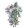
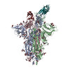
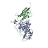
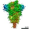
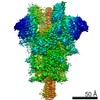
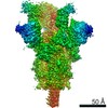
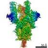
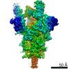
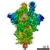
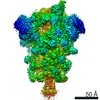
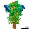
全体 : Spike protein (S-6P, 2-up) in complex with Synthetic nanobody (Sb45)
全体
名称: Spike protein (S-6P, 2-up) in complex with Synthetic nanobody (Sb45)
要素
複合体: Spike protein (S-6P, 2-up) in complex with Synthetic nanobody (Sb45)
タンパク質・ペプチド: Spike glycoprotein
タンパク質・ペプチド: Synthetic nanobody (Sb45)
リガンド: 2-acetamido-2-deoxy-beta-D-glucopyranose
-
超分子 #1: Spike protein (S-6P, 2-up) in complex with Synthetic nanobody (Sb45)
超分子
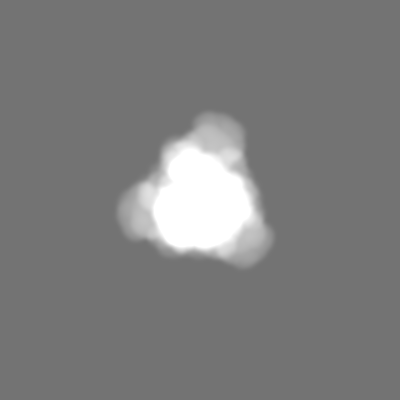
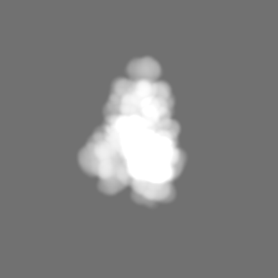
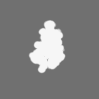
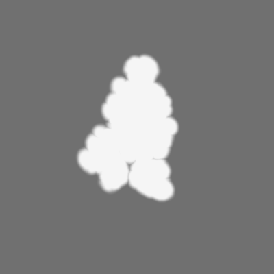
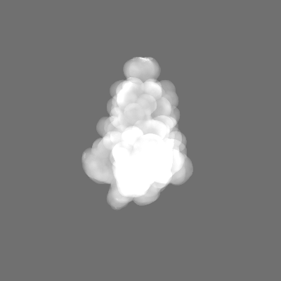
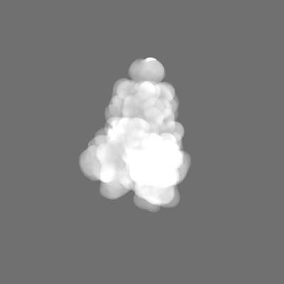
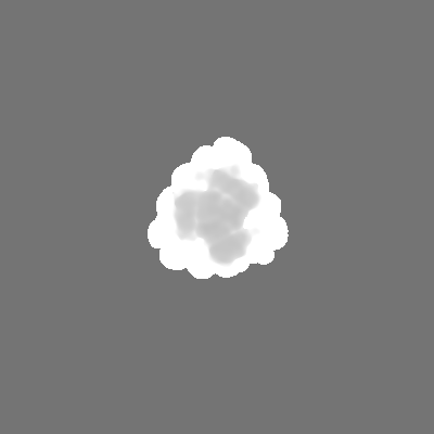
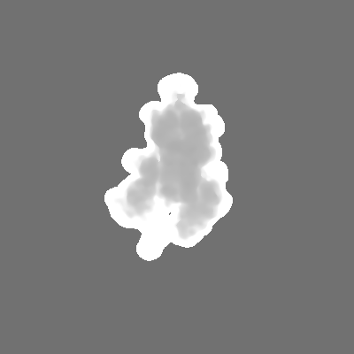
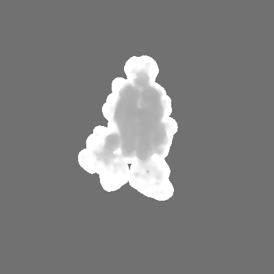
名称: Spike protein (S-6P, 2-up) in complex with Synthetic nanobody (Sb45) タイプ: complex / ID: 1 / 親要素: 0 / 含まれる分子: #1-#2 詳細: Synthetic nanobody Sb45 was mixed with freshly purified S-6P in the mole ratio of 3:1, incubated, and subjected to Negative stain and frozen grids for cryoEM data collection.
選択した数: 1433963 詳細: A total of 9,725 micrographs were imported into cryoSPARC. Following patch Motion correction, patch CTF estimation, and curation, the number of micrographs was reduced to 9,237. The blob ...詳細: A total of 9,725 micrographs were imported into cryoSPARC. Following patch Motion correction, patch CTF estimation, and curation, the number of micrographs was reduced to 9,237. The blob picker with the particle diameter between 128 and 256 angstroms was used for picking up particles. We used the box size of 400 pixels and extracted 1,433,963 particles initially.
詳細: The initial model was built based on 6XKL, but RBD and Sb45 are from 7GKJ and dock on S-6P (6XKL). NTD was replaced by 7B32 with a full sequence.
最終 再構成
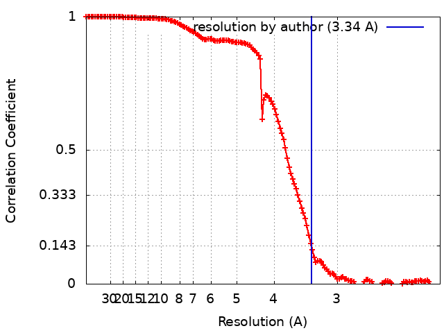
使用したクラス数: 18 / 想定した対称性 - 点群: C1 (非対称) / アルゴリズム: SIMULTANEOUS ITERATIVE (SIRT) / 解像度のタイプ: BY AUTHOR / 解像度: 3.34 Å / 解像度の算出法: FSC 0.143 CUT-OFF / ソフトウェア - 名称: cryoSPARC (ver. V3.2.0) 詳細: A series of Ab-initio 3D reconstruction (classification)dividing 2 or 4 subclasses to identify two forms of S-6P, i.e. one RBD up, or two RBD up. 使用した粒子像数: 61026
初期 角度割当
タイプ: NOT APPLICABLE
最終 角度割当
タイプ: NOT APPLICABLE
最終 3次元分類
クラス数: 100 / ソフトウェア - 名称: cryoSPARC (ver. 3.2.0) 詳細: The best 18 classes were selected with 662,994 particles. 3D classifications further identify two conformations of S-6P: 1-up RBD with 214,171 particles, 2-up RBD with 61,026 particles
An initial model for S-6P was generated using PDB 6XKL and rigid body fitted into the map using Chimera. The RBD domain (334-528) and Sb45 are taken from 7KGJ which was superimposed onto the S-6P model in PyMol. The NTD domain (14-289) is taken from 7B32 with full sequence and replace that of 6XKL. We have rebuilt and added more glycans (NAGs). We used the real-space refinement in PHENIX including rigid-body refinement. RBD and NTD are subjected to rigid-body refinement. Simulate annealing (SA) runs once at the initial micro-step, local grid search and ADP refinements were included, using the secondary structure restraints.
精密化
空間: REAL / プロトコル: RIGID BODY FIT / 温度因子: 157 / 当てはまり具合の基準: Correlation Coefficient
得られたモデル
PDB-7n0h: CryoEM structure of SARS-CoV-2 spike protein (S-6P, 2-up) in complex with sybodies (Sb45)
 ムービー
ムービー コントローラー


 データを開く
 基本情報
基本情報 マップデータ
 試料
試料 機能・相同性情報
 Severe acute respiratory syndrome coronavirus 2 (ウイルス)
 データ登録者
 引用
引用
 構造の表示
構造の表示
 ダウンロードとリンク
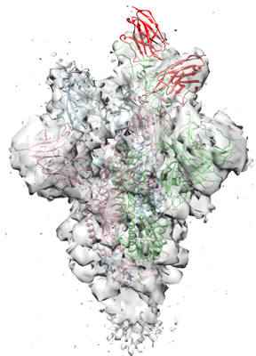 emd_24106.png
 http://ftp.pdbj.org/pub/emdb/structures/EMD-24106


 Z
Z Y
Y X
X


 試料の構成要素
 Homo sapiens (ヒト)

 解析
解析 電子顕微鏡法
 FIELD EMISSION GUN