[English] 日本語
 Yorodumi
- PDB-7n0g: CryoEm structure of SARS-CoV-2 spike protein (S-6P, 1-up) in comp... -
+ Open data
- Basic information
| Entry | Database: PDB / ID: 7n0g | ||||||
|---|---|---|---|---|---|---|---|
| Title | CryoEm structure of SARS-CoV-2 spike protein (S-6P, 1-up) in complex with sybodies (Sb45) | ||||||
 Components Components |
| ||||||
 Keywords Keywords | VIRAL PROTEIN / SARS-CoV-2 / Spike Protein / S-6P (HexaPro) / nanobody / sybody / Sb45 / neutralization | ||||||
| Function / homology |  Function and homology information Function and homology informationsymbiont-mediated disruption of host tissue / Maturation of spike protein / Translation of Structural Proteins / Virion Assembly and Release / host cell surface / host extracellular space / viral translation / symbiont-mediated-mediated suppression of host tetherin activity / Induction of Cell-Cell Fusion / structural constituent of virion ...symbiont-mediated disruption of host tissue / Maturation of spike protein / Translation of Structural Proteins / Virion Assembly and Release / host cell surface / host extracellular space / viral translation / symbiont-mediated-mediated suppression of host tetherin activity / Induction of Cell-Cell Fusion / structural constituent of virion / membrane fusion / entry receptor-mediated virion attachment to host cell / Attachment and Entry / host cell endoplasmic reticulum-Golgi intermediate compartment membrane / positive regulation of viral entry into host cell / receptor-mediated virion attachment to host cell / host cell surface receptor binding / symbiont-mediated suppression of host innate immune response / receptor ligand activity / endocytosis involved in viral entry into host cell / fusion of virus membrane with host plasma membrane / fusion of virus membrane with host endosome membrane / viral envelope / symbiont entry into host cell / virion attachment to host cell / SARS-CoV-2 activates/modulates innate and adaptive immune responses / host cell plasma membrane / virion membrane / identical protein binding / membrane / plasma membrane Similarity search - Function | ||||||
| Biological species |  synthetic construct (others) | ||||||
| Method | ELECTRON MICROSCOPY / single particle reconstruction / cryo EM / Resolution: 3.02 Å | ||||||
 Authors Authors | Jiang, J. / Huang, R. / Margulies, D. | ||||||
 Citation Citation |  Journal: J Biol Chem / Year: 2021 Journal: J Biol Chem / Year: 2021Title: Structures of synthetic nanobody-SARS-CoV-2 receptor-binding domain complexes reveal distinct sites of interaction. Authors: Javeed Ahmad / Jiansheng Jiang / Lisa F Boyd / Allison Zeher / Rick Huang / Di Xia / Kannan Natarajan / David H Margulies /  Abstract: Combating the worldwide spread of severe acute respiratory syndrome coronavirus 2 (SARS-CoV-2) and the emergence of new variants demands understanding of the structural basis of the interaction of ...Combating the worldwide spread of severe acute respiratory syndrome coronavirus 2 (SARS-CoV-2) and the emergence of new variants demands understanding of the structural basis of the interaction of antibodies with the SARS-CoV-2 receptor-binding domain (RBD). Here, we report five X-ray crystal structures of sybodies (synthetic nanobodies) including those of binary and ternary complexes of Sb16-RBD, Sb45-RBD, Sb14-RBD-Sb68, and Sb45-RBD-Sb68, as well as unliganded Sb16. These structures reveal that Sb14, Sb16, and Sb45 bind the RBD at the angiotensin-converting enzyme 2 interface and that the Sb16 interaction is accompanied by a large conformational adjustment of complementarity-determining region 2. In contrast, Sb68 interacts at the periphery of the SARS-CoV-2 RBD-angiotensin-converting enzyme 2 interface. We also determined cryo-EM structures of Sb45 bound to the SARS-CoV-2 spike protein. Superposition of the X-ray structures of sybodies onto the trimeric spike protein cryo-EM map indicates that some sybodies may bind in both "up" and "down" configurations, but others may not. Differences in sybody recognition of several recently identified RBD variants are explained by these structures. | ||||||
| History |
|
- Structure visualization
| Movie |
 Movie viewer Movie viewer |
|---|---|
| Structure viewer | Molecule:  Molmil Molmil Jmol/JSmol Jmol/JSmol |
- Downloads & links
- Download
| PDBx/mmCIF format |  7n0g.cif.gz 7n0g.cif.gz | 655.8 KB | Display |  PDBx/mmCIF format PDBx/mmCIF format |
|---|---|---|---|---|
| PDB format |  pdb7n0g.ent.gz pdb7n0g.ent.gz | 537.8 KB | Display |  PDB format PDB format |
| PDBx/mmJSON format |  7n0g.json.gz 7n0g.json.gz | Tree view |  PDBx/mmJSON format PDBx/mmJSON format | |
| Others |  Other downloads Other downloads |
-Validation report
| Arichive directory |  https://data.pdbj.org/pub/pdb/validation_reports/n0/7n0g https://data.pdbj.org/pub/pdb/validation_reports/n0/7n0g ftp://data.pdbj.org/pub/pdb/validation_reports/n0/7n0g ftp://data.pdbj.org/pub/pdb/validation_reports/n0/7n0g | HTTPS FTP |
|---|
-Related structure data
| Related structure data |  24105MC 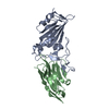 7kgjC 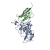 7kgkC 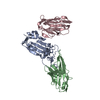 7klwC 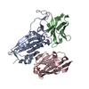 7mfuC 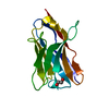 7mfvC  7n0hC C: citing same article ( M: map data used to model this data |
|---|---|
| Similar structure data |
- Links
Links
- Assembly
| Deposited unit | 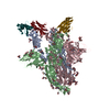
|
|---|---|
| 1 |
|
- Components
| #1: Protein | Mass: 142427.438 Da / Num. of mol.: 3 Source method: isolated from a genetically manipulated source Source: (gene. exp.)  Gene: S, 2 / Cell line (production host): HEK293 / Production host:  Homo sapiens (human) / References: UniProt: P0DTC2 Homo sapiens (human) / References: UniProt: P0DTC2#2: Antibody | Mass: 13574.048 Da / Num. of mol.: 3 Source method: isolated from a genetically manipulated source Source: (gene. exp.) synthetic construct (others) / Plasmid: pET21b / Production host:  #3: Polysaccharide | 2-acetamido-2-deoxy-beta-D-glucopyranose-(1-4)-2-acetamido-2-deoxy-beta-D-glucopyranose Source method: isolated from a genetically manipulated source #4: Sugar | ChemComp-NAG / Has ligand of interest | Y | Has protein modification | Y | |
|---|
-Experimental details
-Experiment
| Experiment | Method: ELECTRON MICROSCOPY |
|---|---|
| EM experiment | Aggregation state: PARTICLE / 3D reconstruction method: single particle reconstruction |
- Sample preparation
| Component | Name: Spike protein (S-6P) in complex with Synthetic nanobody (Sb45) Type: COMPLEX Details: Synthetic nanobody Sb45 was mixed with freshly purified S-6P in the mole ratio of 3:1, incubated, and subjected to Negative stain and frozen grids for cryoEM data collection. Entity ID: #1-#2 / Source: RECOMBINANT | ||||||||||||
|---|---|---|---|---|---|---|---|---|---|---|---|---|---|
| Molecular weight | Value: 0.6 MDa / Experimental value: NO | ||||||||||||
| Source (natural) |
| ||||||||||||
| Source (recombinant) |
| ||||||||||||
| Buffer solution | pH: 8 | ||||||||||||
| Buffer component | Conc.: 0.25 mg/mL / Name: TBS | ||||||||||||
| Specimen | Conc.: 0.6 mg/ml / Embedding applied: NO / Shadowing applied: NO / Staining applied: NO / Vitrification applied: YES | ||||||||||||
| Specimen support | Grid material: COPPER / Grid mesh size: 300 divisions/in. / Grid type: Quantifoil R1.2/1.3 | ||||||||||||
| Vitrification | Instrument: FEI VITROBOT MARK IV / Cryogen name: ETHANE / Humidity: 95 % / Chamber temperature: 277 K |
- Electron microscopy imaging
| Experimental equipment |  Model: Titan Krios / Image courtesy: FEI Company |
|---|---|
| Microscopy | Model: FEI TITAN KRIOS |
| Electron gun | Electron source:  FIELD EMISSION GUN / Accelerating voltage: 300 kV / Illumination mode: FLOOD BEAM FIELD EMISSION GUN / Accelerating voltage: 300 kV / Illumination mode: FLOOD BEAM |
| Electron lens | Mode: BRIGHT FIELD / Nominal magnification: 130000 X / Calibrated magnification: 47529 X / Nominal defocus max: 2000 nm / Nominal defocus min: 700 nm / Calibrated defocus min: 535 nm / Calibrated defocus max: 2431 nm / Cs: 2.7 mm / C2 aperture diameter: 100 µm / Alignment procedure: BASIC |
| Specimen holder | Cryogen: NITROGEN / Specimen holder model: FEI TITAN KRIOS AUTOGRID HOLDER / Temperature (max): 80 K / Temperature (min): 78 K |
| Image recording | Average exposure time: 8 sec. / Electron dose: 56 e/Å2 / Detector mode: SUPER-RESOLUTION / Film or detector model: GATAN K2 QUANTUM (4k x 4k) / Num. of grids imaged: 2 / Num. of real images: 9725 |
| Image scans | Sampling size: 5.001 µm / Movie frames/image: 40 |
- Processing
| Software | Name: PHENIX / Version: 1.19.2_4158: / Classification: refinement | ||||||||||||||||||||||||||||||||||||||||||
|---|---|---|---|---|---|---|---|---|---|---|---|---|---|---|---|---|---|---|---|---|---|---|---|---|---|---|---|---|---|---|---|---|---|---|---|---|---|---|---|---|---|---|---|
| EM software |
| ||||||||||||||||||||||||||||||||||||||||||
| CTF correction | Details: patch CTF estimation / Type: NONE | ||||||||||||||||||||||||||||||||||||||||||
| Particle selection | Num. of particles selected: 1433963 Details: A total of 9,725 micrographs were imported into cryoSPARC. Following patch Motion correction, patch CTF estimation, and curation, the number of micrographs was reduced to 9,237. The blob ...Details: A total of 9,725 micrographs were imported into cryoSPARC. Following patch Motion correction, patch CTF estimation, and curation, the number of micrographs was reduced to 9,237. The blob picker with the particle diameter between 128 and 256 angstroms was used for picking up particles. We used the box size of 400 pixels and extracted 1,433,963 particles initially. | ||||||||||||||||||||||||||||||||||||||||||
| Symmetry | Point symmetry: C1 (asymmetric) | ||||||||||||||||||||||||||||||||||||||||||
| 3D reconstruction | Resolution: 3.02 Å / Resolution method: FSC 0.143 CUT-OFF / Num. of particles: 214171 / Algorithm: SIMULTANEOUS ITERATIVE (SIRT) Details: A series of "Ab-initio 3D reconstruction" dividing 2 or 4 subclasses to identify two forms of configurations of S-6P, i.e. one RBD up or two RBD up. Num. of class averages: 18 / Symmetry type: POINT | ||||||||||||||||||||||||||||||||||||||||||
| Atomic model building | B value: 157 / Protocol: RIGID BODY FIT / Space: REAL / Target criteria: Correlation Coefficient Details: An initial model for S-6P was generated using PDB 6XKL and rigid body fitted into the map using Chimera. The RBD domain (334-528) and Sb45 are taken from 7KGJ which was superimposed onto the ...Details: An initial model for S-6P was generated using PDB 6XKL and rigid body fitted into the map using Chimera. The RBD domain (334-528) and Sb45 are taken from 7KGJ which was superimposed onto the S-6P model in PyMol. The NTD domain (14-289) is taken from 7B32 with full sequence and replace that of 6XKL. We have rebuilt and added more glycans (NAGs). We used the real-space refinement in PHENIX including rigid-body refinement. RBD and NTD are subjected to rigid-body refinement. Simulate annealing (SA) runs once at the initial micro-step, local grid search and ADP refinements were included, using the secondary structure restraints. | ||||||||||||||||||||||||||||||||||||||||||
| Atomic model building | 3D fitting-ID: 1 / Source name: PDB / Type: experimental model
| ||||||||||||||||||||||||||||||||||||||||||
| Refine LS restraints |
|
 Movie
Movie Controller






 PDBj
PDBj


