ムービー
ムービー コントローラー

[日本語] English
 万見
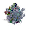
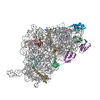
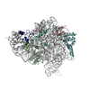
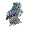
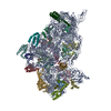
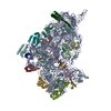
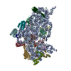
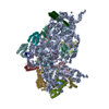
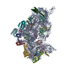
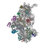
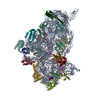
万見- EMDB-23052: Staphylococcus aureus 30S ribosomal subunit in presence of spermidine -
+ データを開く

- 基本情報
基本情報
| 登録情報 | データベース: EMDB / ID: EMD-23052 | |||||||||
|---|---|---|---|---|---|---|---|---|---|---|
| タイトル | Staphylococcus aureus 30S ribosomal subunit in presence of spermidine | |||||||||
 マップデータ マップデータ | ||||||||||
 試料 試料 |
| |||||||||
 キーワード キーワード | Pathogen / small ribosomal subunit / spermidine / RIBOSOME | |||||||||
| 機能・相同性 |  機能・相同性情報 機能・相同性情報ribosomal small subunit biogenesis / ribosomal small subunit assembly / small ribosomal subunit / small ribosomal subunit rRNA binding / cytosolic small ribosomal subunit / tRNA binding / rRNA binding / ribosome / structural constituent of ribosome / translation ...ribosomal small subunit biogenesis / ribosomal small subunit assembly / small ribosomal subunit / small ribosomal subunit rRNA binding / cytosolic small ribosomal subunit / tRNA binding / rRNA binding / ribosome / structural constituent of ribosome / translation / mRNA binding / RNA binding / zinc ion binding / cytoplasm / cytosol 類似検索 - 分子機能 | |||||||||
| 生物種 |  Staphylococcus aureus (strain NCTC 8325 / PS 47) (黄色ブドウ球菌) / Staphylococcus aureus subsp. aureus NCTC 8325 (黄色ブドウ球菌) Staphylococcus aureus (strain NCTC 8325 / PS 47) (黄色ブドウ球菌) / Staphylococcus aureus subsp. aureus NCTC 8325 (黄色ブドウ球菌) | |||||||||
| 手法 | 単粒子再構成法 / クライオ電子顕微鏡法 / 解像度: 3.75 Å | |||||||||
 データ登録者 データ登録者 | Belinite M / Khusainov I | |||||||||
| 資金援助 |  フランス, 2件 フランス, 2件
| |||||||||
 引用 引用 | ジャーナル: Front Mol Biosci / 年: 2021 タイトル: Stabilization of Ribosomal RNA of the Small Subunit by Spermidine in . 著者: Margarita Belinite / Iskander Khusainov / Heddy Soufari / Stefano Marzi / Pascale Romby / Marat Yusupov / Yaser Hashem /  要旨: Cryo-electron microscopy is now used as a method of choice in structural biology for studying protein synthesis, a process mediated by the ribosome machinery. In order to achieve high-resolution ...Cryo-electron microscopy is now used as a method of choice in structural biology for studying protein synthesis, a process mediated by the ribosome machinery. In order to achieve high-resolution structures using this approach, one needs to obtain homogeneous and stable samples, which requires optimization of ribosome purification in a species-dependent manner. This is especially critical for the bacterial small ribosomal subunit that tends to be unstable in the absence of ligands. Here, we report a protocol for purification of stable 30 S from the Gram-positive bacterium and its cryo-EM structures: in presence of spermidine at a resolution ranging between 3.4 and 3.6 Å and in its absence at 5.3 Å. Using biochemical characterization and cryo-EM, we demonstrate the importance of spermidine for stabilization of the 30 S preserving favorable conformation of the helix 44. | |||||||||
| 履歴 |
|
- 構造の表示
構造の表示
| ムービー |
ムービービューア |
|---|---|
| 構造ビューア | EMマップ: SurfViewMolmilJmol/JSmol |
| 添付画像 |

- ダウンロードとリンク
-EMDBアーカイブ
| マップデータ | emd_23052.map.gz | 70.1 MB | EMDBマップデータ形式 | |
|---|---|---|---|---|
| ヘッダ (付随情報) | emd-23052-v30.xmlemd-23052.xml | 31.2 KB 31.2 KB | 表示 表示 | EMDBヘッダ |
| 画像 | 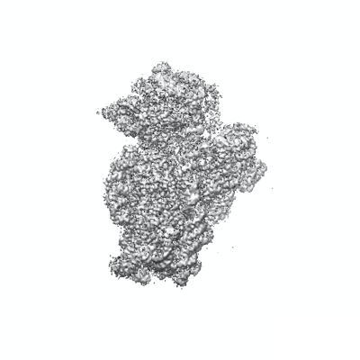 emd_23052.png emd_23052.png | 90 KB | ||
| Filedesc metadata | emd-23052.cif.gz | 8.1 KB | ||
| アーカイブディレクトリ |  http://ftp.pdbj.org/pub/emdb/structures/EMD-23052ftp://ftp.pdbj.org/pub/emdb/structures/EMD-23052 http://ftp.pdbj.org/pub/emdb/structures/EMD-23052ftp://ftp.pdbj.org/pub/emdb/structures/EMD-23052 | HTTPS FTP |
-検証レポート
| 文書・要旨 | emd_23052_validation.pdf.gz | 562 KB | 表示 | EMDB検証レポート |
|---|---|---|---|---|
| 文書・詳細版 | emd_23052_full_validation.pdf.gz | 561.5 KB | 表示 | |
| XML形式データ | emd_23052_validation.xml.gz | 6.2 KB | 表示 | |
| CIF形式データ | emd_23052_validation.cif.gz | 7.1 KB | 表示 | |
| アーカイブディレクトリ | https://ftp.pdbj.org/pub/emdb/validation_reports/EMD-23052ftp://ftp.pdbj.org/pub/emdb/validation_reports/EMD-23052 | HTTPS FTP |
-関連構造データ


-リンク
| EMDBのページ | EMDB (EBI/PDBe) / EMDataResource |
|---|---|
| 「今月の分子」の関連する項目 |


-マップ
| ファイル | ダウンロード / ファイル: emd_23052.map.gz / 形式: CCP4 / 大きさ: 75.1 MB / タイプ: IMAGE STORED AS FLOATING POINT NUMBER (4 BYTES) | ||||||||||||||||||||||||||||||||||||||||||||||||||||||||||||
|---|---|---|---|---|---|---|---|---|---|---|---|---|---|---|---|---|---|---|---|---|---|---|---|---|---|---|---|---|---|---|---|---|---|---|---|---|---|---|---|---|---|---|---|---|---|---|---|---|---|---|---|---|---|---|---|---|---|---|---|---|---|
| ボクセルのサイズ | X=Y=Z: 1.19 Å | ||||||||||||||||||||||||||||||||||||||||||||||||||||||||||||
| 密度 |
| ||||||||||||||||||||||||||||||||||||||||||||||||||||||||||||
| 対称性 | 空間群: 1 | ||||||||||||||||||||||||||||||||||||||||||||||||||||||||||||
| 詳細 | EMDB XML:
CCP4マップ ヘッダ情報:
| ||||||||||||||||||||||||||||||||||||||||||||||||||||||||||||
-添付データ
- 試料の構成要素
+全体 : Staphylococcus aureus 30S ribosomal subunit in presence of spermidine
+超分子 #1: Staphylococcus aureus 30S ribosomal subunit in presence of spermidine
+分子 #1: mRNA
+分子 #2: 16S ribosomal RNA
+分子 #3: 30S ribosomal protein S2
+分子 #4: 30S ribosomal protein S3
+分子 #5: 30S ribosomal protein S4
+分子 #6: 30S ribosomal protein S5
+分子 #7: 30S ribosomal protein S6
+分子 #8: 30S ribosomal protein S7
+分子 #9: 30S ribosomal protein S8
+分子 #10: 30S ribosomal protein S9
+分子 #11: 30S ribosomal protein S10
+分子 #12: 30S ribosomal protein S11
+分子 #13: 30S ribosomal protein S12
+分子 #14: 30S ribosomal protein S13
+分子 #15: 30S ribosomal protein S14 type Z
+分子 #16: 30S ribosomal protein S15
+分子 #17: 30S ribosomal protein S16
+分子 #18: 30S ribosomal protein S17
+分子 #19: 30S ribosomal protein S18
+分子 #20: 30S ribosomal protein S19
+分子 #21: 30S ribosomal protein S20
-実験情報
-構造解析
| 手法 | クライオ電子顕微鏡法 |
|---|---|
 解析 解析 | 単粒子再構成法 |
| 試料の集合状態 | particle |
-試料調製
| 緩衝液 | pH: 7.5 |
|---|---|
| 凍結 | 凍結剤: ETHANE / 装置: FEI VITROBOT MARK IV |
- 電子顕微鏡法
| 顕微鏡 | FEI TALOS ARCTICA |
|---|---|
| 撮影 | フィルム・検出器のモデル: FEI FALCON III (4k x 4k) 平均電子線量: 3.0 e/Å2 |
| 電子線 | 加速電圧: 200 kV / 電子線源:  FIELD EMISSION GUN FIELD EMISSION GUN |
| 電子光学系 | 照射モード: OTHER / 撮影モード: BRIGHT FIELD |
| 実験機器 |  モデル: Talos Arctica / 画像提供: FEI Company |
-画像解析
| 初期モデル | モデルのタイプ: PDB ENTRY PDBモデル - PDB ID: |
|---|---|
| 最終 再構成 | 解像度のタイプ: BY AUTHOR / 解像度: 3.75 Å / 解像度の算出法: FSC 0.143 CUT-OFF / 使用した粒子像数: 529602 |
| 初期 角度割当 | タイプ: PROJECTION MATCHING |
| 最終 角度割当 | タイプ: PROJECTION MATCHING |