National Institutes of Health/National Institute Of Allergy and Infectious Diseases (NIH/NIAID)
P01-AI138398-S1
United States
Citation
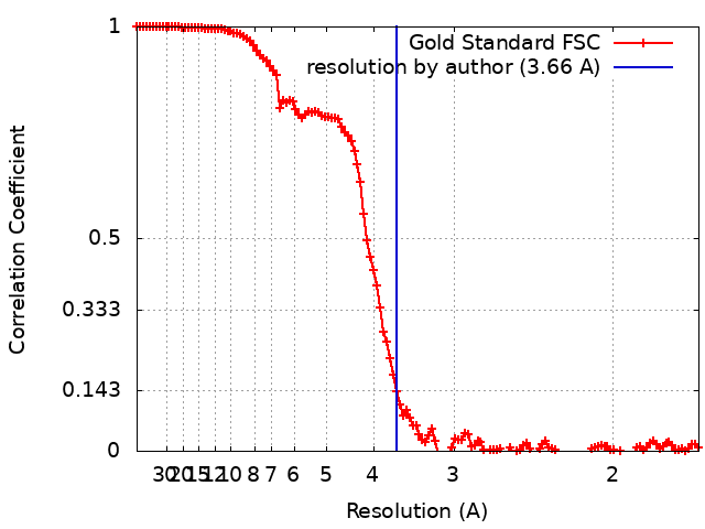
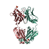
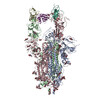
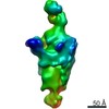
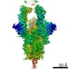
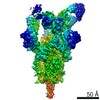
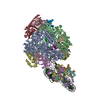
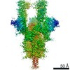
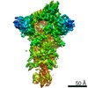
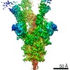
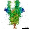
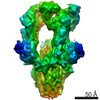
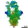
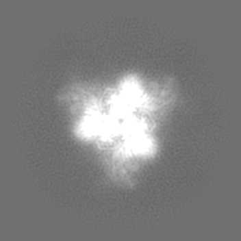
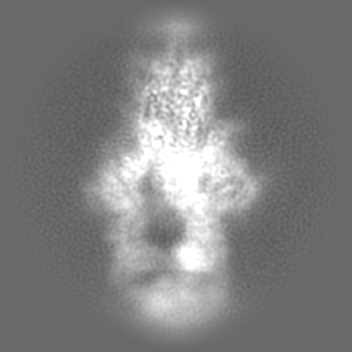
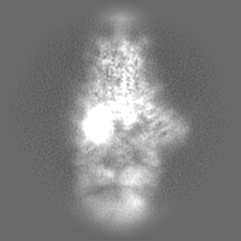
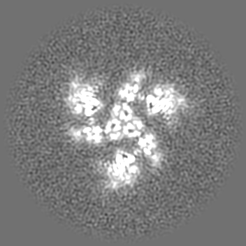
Journal: Cell / Year: 2020 Title: Structures of Human Antibodies Bound to SARS-CoV-2 Spike Reveal Common Epitopes and Recurrent Features of Antibodies. Authors: Christopher O Barnes / Anthony P West / Kathryn E Huey-Tubman / Magnus A G Hoffmann / Naima G Sharaf / Pauline R Hoffman / Nicholas Koranda / Harry B Gristick / Christian Gaebler / Frauke ...Authors: Christopher O Barnes / Anthony P West / Kathryn E Huey-Tubman / Magnus A G Hoffmann / Naima G Sharaf / Pauline R Hoffman / Nicholas Koranda / Harry B Gristick / Christian Gaebler / Frauke Muecksch / Julio C Cetrulo Lorenzi / Shlomo Finkin / Thomas Hägglöf / Arlene Hurley / Katrina G Millard / Yiska Weisblum / Fabian Schmidt / Theodora Hatziioannou / Paul D Bieniasz / Marina Caskey / Davide F Robbiani / Michel C Nussenzweig / Pamela J Bjorkman / Abstract: Neutralizing antibody responses to coronaviruses mainly target the receptor-binding domain (RBD) of the trimeric spike. Here, we characterized polyclonal immunoglobulin Gs (IgGs) and Fabs from COVID- ...Neutralizing antibody responses to coronaviruses mainly target the receptor-binding domain (RBD) of the trimeric spike. Here, we characterized polyclonal immunoglobulin Gs (IgGs) and Fabs from COVID-19 convalescent individuals for recognition of coronavirus spikes. Plasma IgGs differed in their focus on RBD epitopes, recognition of alpha- and beta-coronaviruses, and contributions of avidity to increased binding/neutralization of IgGs over Fabs. Using electron microscopy, we examined specificities of polyclonal plasma Fabs, revealing recognition of both S1 and RBD epitopes on SARS-CoV-2 spike. Moreover, a 3.4 Å cryo-electron microscopy (cryo-EM) structure of a neutralizing monoclonal Fab-spike complex revealed an epitope that blocks ACE2 receptor binding. Modeling based on these structures suggested different potentials for inter-spike crosslinking by IgGs on viruses, and characterized IgGs would not be affected by identified SARS-CoV-2 spike mutations. Overall, our studies structurally define a recurrent anti-SARS-CoV-2 antibody class derived from VH3-53/VH3-66 and similarity to a SARS-CoV VH3-30 antibody, providing criteria for evaluating vaccine-elicited antibodies.
History
Deposition
Jun 8, 2020
-
Header (metadata) release
Jul 1, 2020
-
Map release
Jul 1, 2020
-
Update
Nov 6, 2024
-
Current status
Nov 6, 2024
Processing site: RCSB / Status: Released
-
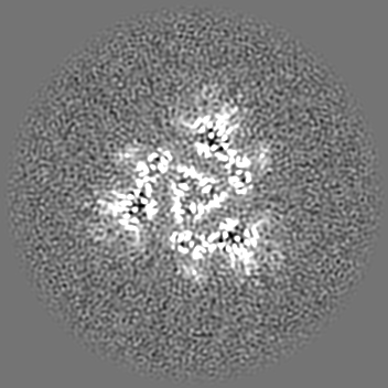
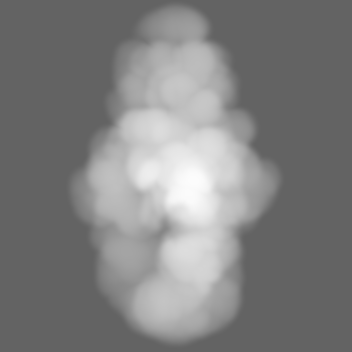
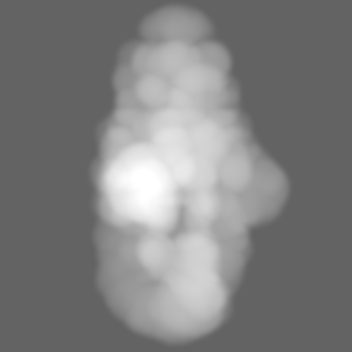
Structure visualization
Movie
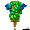
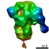
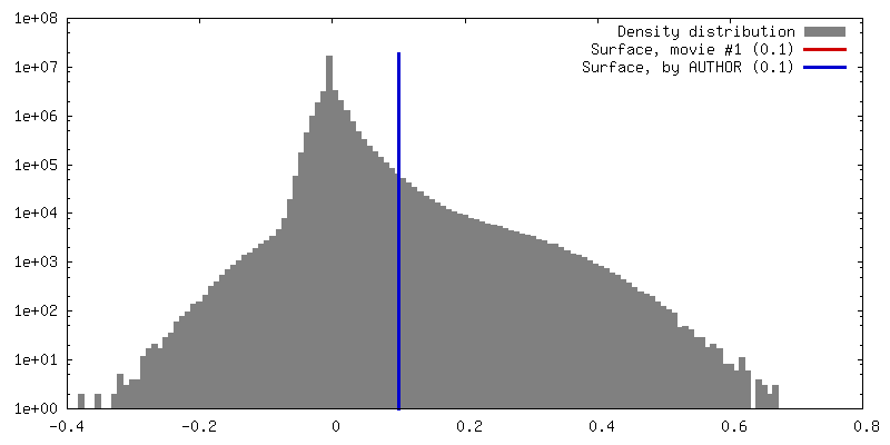
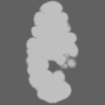
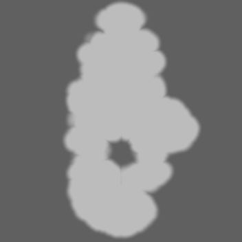
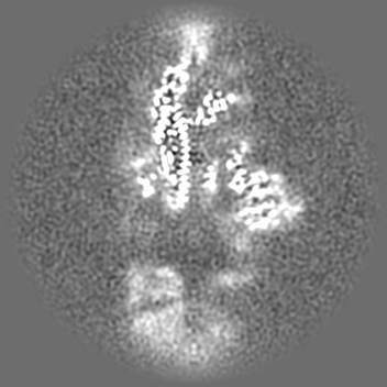
Surface view with section colored by density value
Model: UltrAuFoil R1.2/1.3 / Material: GOLD / Mesh: 300 / Support film - Material: GOLD / Support film - topology: HOLEY ARRAY / Pretreatment - Type: GLOW DISCHARGE / Pretreatment - Time: 60 sec. / Pretreatment - Atmosphere: AIR / Details: 60 s glow discharge in air at 0.20 mA current
Vitrification
Cryogen name: ETHANE / Chamber humidity: 100 % / Chamber temperature: 295 K / Instrument: FEI VITROBOT MARK IV Details: 3 microliters added, blotted for 3 s with 0 blot force.
Details
Monodisperse sample taken after size-exclusion chromatography
-
Electron microscopy
Microscope
FEI TITAN KRIOS
Details
Collected with 3x3 pattern and beam image shift
Image recording
Film or detector model: GATAN K3 BIOQUANTUM (6k x 4k) / Number grids imaged: 1 / Number real images: 5940 / Average exposure time: 1.89 sec. / Average electron dose: 60.0 e/Å2
Electron beam
Acceleration voltage: 300 kV / Electron source: FIELD EMISSION GUN
In the structure databanks used in Yorodumi, some data are registered as the other names, "COVID-19 virus" and "2019-nCoV". Here are the details of the virus and the list of structure data.
Jan 31, 2019. EMDB accession codes are about to change! (news from PDBe EMDB page)
EMDB accession codes are about to change! (news from PDBe EMDB page)
The allocation of 4 digits for EMDB accession codes will soon come to an end. Whilst these codes will remain in use, new EMDB accession codes will include an additional digit and will expand incrementally as the available range of codes is exhausted. The current 4-digit format prefixed with “EMD-” (i.e. EMD-XXXX) will advance to a 5-digit format (i.e. EMD-XXXXX), and so on. It is currently estimated that the 4-digit codes will be depleted around Spring 2019, at which point the 5-digit format will come into force.
The EM Navigator/Yorodumi systems omit the EMD- prefix.
Related info.:Q: What is EMD? / ID/Accession-code notation in Yorodumi/EM Navigator
Yorodumi is a browser for structure data from EMDB, PDB, SASBDB, etc.
This page is also the successor to EM Navigator detail page, and also detail information page/front-end page for Omokage search.
The word "yorodu" (or yorozu) is an old Japanese word meaning "ten thousand". "mi" (miru) is to see.
Related info.:EMDB / PDB / SASBDB / Comparison of 3 databanks / Yorodumi Search / Aug 31, 2016. New EM Navigator & Yorodumi / Yorodumi Papers / Jmol/JSmol / Function and homology information / Changes in new EM Navigator and Yorodumi
 Movie
Movie Controller

 Yorodumi
 Open data
 Basic information
 Map data
 Sample
 Keywords
 Function and homology information
 Severe acute respiratory syndrome coronavirus 2 /
  Homo sapiens (human)
 Authors
 United States, 1 items
  Citation
 Structure visualization
 Downloads & links
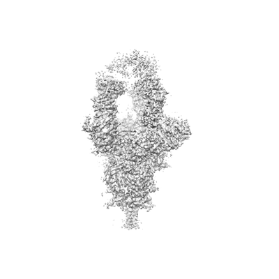 emd_22128.png
 http://ftp.pdbj.org/pub/emdb/structures/EMD-22128


 Z (Sec.)
 Y (Row.)
 X (Col.)


 Sample components
 Processing
 Electron microscopy
 FIELD EMISSION GUN