+ Open data
- Basic information
| Entry | Database: EMDB / ID: EMD-21095 | ||||||||||||||||||
|---|---|---|---|---|---|---|---|---|---|---|---|---|---|---|---|---|---|---|---|
| Title | Parainfluenza virus 5 L-P complex | ||||||||||||||||||
 Map data Map data | |||||||||||||||||||
 Sample Sample |
| ||||||||||||||||||
 Keywords Keywords | VIRAL PROTEIN / POLYMERASE / METHYLTRANSFERASE / POLY-RIBONUCLEOTIDYLTRANSFERASE | ||||||||||||||||||
| Function / homology |  Function and homology information Function and homology informationNNS virus cap methyltransferase / GDP polyribonucleotidyltransferase / Hydrolases; Acting on acid anhydrides; In phosphorus-containing anhydrides / virion component / mRNA 5'-cap (guanine-N7-)-methyltransferase activity / host cell cytoplasm / RNA-directed RNA polymerase / RNA-dependent RNA polymerase activity / GTPase activity / ATP binding Similarity search - Function | ||||||||||||||||||
| Biological species |  Simian virus 5 (strain W3) / Simian virus 5 (strain W3) /  Parainfluenza virus 5 (strain W3) Parainfluenza virus 5 (strain W3) | ||||||||||||||||||
| Method | single particle reconstruction / cryo EM / Resolution: 4.38 Å | ||||||||||||||||||
 Authors Authors | Abdella R / He Y | ||||||||||||||||||
| Funding support |  United States, 5 items United States, 5 items
| ||||||||||||||||||
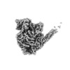
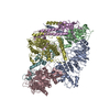
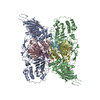
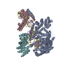
 Citation Citation |  Journal: Proc Natl Acad Sci U S A / Year: 2020 Journal: Proc Natl Acad Sci U S A / Year: 2020Title: Structure of a paramyxovirus polymerase complex reveals a unique methyltransferase-CTD conformation. Authors: Ryan Abdella / Megha Aggarwal / Takashi Okura / Robert A Lamb / Yuan He /  Abstract: Paramyxoviruses are enveloped, nonsegmented, negative-strand RNA viruses that cause a wide spectrum of human and animal diseases. The viral genome, packaged by the nucleoprotein (N), serves as a ...Paramyxoviruses are enveloped, nonsegmented, negative-strand RNA viruses that cause a wide spectrum of human and animal diseases. The viral genome, packaged by the nucleoprotein (N), serves as a template for the polymerase complex, composed of the large protein (L) and the homo-tetrameric phosphoprotein (P). The ∼250-kDa L possesses all enzymatic activities necessary for its function but requires P in vivo. Structural information is available for individual P domains from different paramyxoviruses, but how P interacts with L and how that affects the activity of L is largely unknown due to the lack of high-resolution structures of this complex in this viral family. In this study we determined the structure of the L-P complex from parainfluenza virus 5 (PIV5) at 4.3-Å resolution using cryoelectron microscopy, as well as the oligomerization domain (OD) of P at 1.4-Å resolution using X-ray crystallography. P-OD associates with the RNA-dependent RNA polymerase domain of L and protrudes away from it, while the X domain of one chain of P is bound near the L nucleotide entry site. The methyltransferase (MTase) domain and the C-terminal domain (CTD) of L adopt a unique conformation, positioning the MTase active site immediately above the poly-ribonucleotidyltransferase domain and near the likely exit site for the product RNA 5' end. Our study reveals a potential mechanism that mononegavirus polymerases may employ to switch between transcription and genome replication. This knowledge will assist in the design and development of antivirals against paramyxoviruses. | ||||||||||||||||||
| History |
|
- Structure visualization
| Movie |
 Movie viewer Movie viewer |
|---|---|
| Structure viewer | EM map:  SurfView SurfView Molmil Molmil Jmol/JSmol Jmol/JSmol |
| Supplemental images |
- Downloads & links
-EMDB archive
| Map data |  emd_21095.map.gz emd_21095.map.gz | 11.1 MB |  EMDB map data format EMDB map data format | |
|---|---|---|---|---|
| Header (meta data) |  emd-21095-v30.xml emd-21095-v30.xml emd-21095.xml emd-21095.xml | 16.7 KB 16.7 KB | Display Display |  EMDB header EMDB header |
| FSC (resolution estimation) |  emd_21095_fsc.xml emd_21095_fsc.xml | 12.8 KB | Display |  FSC data file FSC data file |
| Images |  emd_21095.png emd_21095.png | 33.4 KB | ||
| Filedesc metadata |  emd-21095.cif.gz emd-21095.cif.gz | 7.3 KB | ||
| Archive directory |  http://ftp.pdbj.org/pub/emdb/structures/EMD-21095 http://ftp.pdbj.org/pub/emdb/structures/EMD-21095 ftp://ftp.pdbj.org/pub/emdb/structures/EMD-21095 ftp://ftp.pdbj.org/pub/emdb/structures/EMD-21095 | HTTPS FTP |
-Validation report
| Summary document |  emd_21095_validation.pdf.gz emd_21095_validation.pdf.gz | 365.4 KB | Display |  EMDB validaton report EMDB validaton report |
|---|---|---|---|---|
| Full document |  emd_21095_full_validation.pdf.gz emd_21095_full_validation.pdf.gz | 364.9 KB | Display | |
| Data in XML |  emd_21095_validation.xml.gz emd_21095_validation.xml.gz | 12.9 KB | Display | |
| Data in CIF |  emd_21095_validation.cif.gz emd_21095_validation.cif.gz | 17.5 KB | Display | |
| Arichive directory |  https://ftp.pdbj.org/pub/emdb/validation_reports/EMD-21095 https://ftp.pdbj.org/pub/emdb/validation_reports/EMD-21095 ftp://ftp.pdbj.org/pub/emdb/validation_reports/EMD-21095 ftp://ftp.pdbj.org/pub/emdb/validation_reports/EMD-21095 | HTTPS FTP |
-Related structure data
| Related structure data |  6v85MC  6v86C  6vagC M: atomic model generated by this map C: citing same article ( |
|---|---|
| Similar structure data |
- Links
Links
| EMDB pages |  EMDB (EBI/PDBe) / EMDB (EBI/PDBe) /  EMDataResource EMDataResource |
|---|---|
| Related items in Molecule of the Month |
- Map
Map
| File |  Download / File: emd_21095.map.gz / Format: CCP4 / Size: 178 MB / Type: IMAGE STORED AS FLOATING POINT NUMBER (4 BYTES) Download / File: emd_21095.map.gz / Format: CCP4 / Size: 178 MB / Type: IMAGE STORED AS FLOATING POINT NUMBER (4 BYTES) | ||||||||||||||||||||||||||||||||||||||||||||||||||||||||||||||||||||
|---|---|---|---|---|---|---|---|---|---|---|---|---|---|---|---|---|---|---|---|---|---|---|---|---|---|---|---|---|---|---|---|---|---|---|---|---|---|---|---|---|---|---|---|---|---|---|---|---|---|---|---|---|---|---|---|---|---|---|---|---|---|---|---|---|---|---|---|---|---|
| Projections & slices | Image control
Images are generated by Spider. | ||||||||||||||||||||||||||||||||||||||||||||||||||||||||||||||||||||
| Voxel size | X=Y=Z: 1.12 Å | ||||||||||||||||||||||||||||||||||||||||||||||||||||||||||||||||||||
| Density |
| ||||||||||||||||||||||||||||||||||||||||||||||||||||||||||||||||||||
| Symmetry | Space group: 1 | ||||||||||||||||||||||||||||||||||||||||||||||||||||||||||||||||||||
| Details | EMDB XML:
CCP4 map header:
| ||||||||||||||||||||||||||||||||||||||||||||||||||||||||||||||||||||
-Supplemental data
- Sample components
-Entire : L-P complex
| Entire | Name: L-P complex |
|---|---|
| Components |
|
-Supramolecule #1: L-P complex
| Supramolecule | Name: L-P complex / type: complex / ID: 1 / Parent: 0 / Macromolecule list: #1-#2 |
|---|---|
| Source (natural) | Organism:  Simian virus 5 (strain W3) Simian virus 5 (strain W3) |
| Molecular weight | Theoretical: 170 KDa |
-Supramolecule #2: L protein
| Supramolecule | Name: L protein / type: complex / ID: 2 / Parent: 1 / Macromolecule list: #1 |
|---|---|
| Source (natural) | Organism:  Simian virus 5 (strain W3) Simian virus 5 (strain W3) |
-Supramolecule #3: P protein
| Supramolecule | Name: P protein / type: complex / ID: 3 / Parent: 1 / Macromolecule list: #2 / Details: homo-tetramer |
|---|---|
| Source (natural) | Organism:  Simian virus 5 (strain W3) Simian virus 5 (strain W3) |
-Macromolecule #1: RNA-directed RNA polymerase L
| Macromolecule | Name: RNA-directed RNA polymerase L / type: protein_or_peptide / ID: 1 / Number of copies: 1 / Enantiomer: LEVO / EC number: RNA-directed RNA polymerase |
|---|---|
| Source (natural) | Organism:  Parainfluenza virus 5 (strain W3) / Strain: W3 Parainfluenza virus 5 (strain W3) / Strain: W3 |
| Molecular weight | Theoretical: 256.195672 KDa |
| Recombinant expression | Organism:  |
| Sequence | String: MAGSREILLP EVHLNSPIVK HKLYYYILLG NLPNEIDLDD LGPLHNQNWN QIAHEESNLA QRLVNVRNFL ITHIPDLRKG HWQEYVNVI LWPRILPLIP DFKINDQLPL LKNWDKLVKE SCSVINAGTS QCIQNLSYGL TGRGNLFTRS RELSGDRRDI D LKTVVAAW ...String: MAGSREILLP EVHLNSPIVK HKLYYYILLG NLPNEIDLDD LGPLHNQNWN QIAHEESNLA QRLVNVRNFL ITHIPDLRKG HWQEYVNVI LWPRILPLIP DFKINDQLPL LKNWDKLVKE SCSVINAGTS QCIQNLSYGL TGRGNLFTRS RELSGDRRDI D LKTVVAAW HDSDWKRISD FWIMIKFQMR QLIVRQTDHN DSDLITYIEN REGIIIITPE LVALFNTENH TLTYMTFEIV LM VSDMYEG RHNILSLCTV STYLNPLKKR ITYLLSLVDN LAFQIGDAVY NIIALLESFV YAQLQMSDPI PELRGQFHAF VCS EILDAL RGTNSFTQDE LRTVTTNLIS PFQDLTPDLT AELLCIMRLW GHPMLTASQA AGKVRESMCA GKVLDFPTIM KTLA FFHTI LINGYRRKHH GVWPPLNLPG NASKGLTELM NDNTEISYEF TLKHWKEVSL IKFKKCFDAD AGEELSIFMK DKAIS APKQ DWMSVFRRSL IKQRHQHHQV PLPNPFNRRL LLNFLGDDKF DPNVELQYVT SGEYLHDDTF CASYSLKEKE IKPDGR IFA KLTKRMRSCQ VIAESLLANH AGKLMKENGV VMNQLSLTKS LLTMSQIGII SEKARKSTRD NINQPGFQNI QRNKSHH SK QVNQRDPSDD FELAASFLTT DLKKYCLQWR YQTIIPFAQS LNRMYGYPHL FEWIHLRLMR STLYVGDPFN PPADTSQF D LDKVINGDIF IVSPRGGIEG LCQKAWTMIS IAVIILSATE SGTRVMSMVQ GDNQAIAVTT RVPRSLPTLE KKTIAFRSC NLFFERLKCN NFGLGHHLKE QETIISSHFF VYSKRIFYQG RILTQALKNA SKLCLTADVL GECTQSSCSN LATTVMRLTE NGVEKDICF YLNIYMTIKQ LSYDIIFPQV SIPGDQITLE YINNPHLVSR LALLPSQLGG LNYLSCSRLF NRNIGDPVVS A VADLKRLI KSGCMDYWIL YNLLGRKPGN GSWATLAADP YSINIEYQYP PTTALKRHTQ QALMELSTNP MLRGIFSDNA QA EENNLAR FLLDREVIFP RVAHIIIEQT SVGRRKQIQG YLDSTRSIMR KSLEIKPLSN RKLNEILDYN INYLAYNLAL LKN AIEPPT YLKAMTLETC SIDIARNLRK LSWAPLLGGR NLEGLETPDP IEITAGALIV GSGYCEQCAA GDNRFTWFFL PSGI EIGGD PRDNPPIRVP YIGSRTDERR VASMAYIRGA SSSLKAVLRL AGVYIWAFGD TLENWIDALD LSHTRVNITL EQLQS LTPL PTSANLTHRL DDGTTTLKFT PASSYTFSSF THISNDEQYL TINDKTADSN IIYQQLMITG LGILETWNNP PINRTF EES TLHLHTGASC CVRPVDSCIL SEALTVKPHI TVPYSNKFVF DEDPLSEYET AKLESLSFQA QLGNIDAVDM TGKLTLL SQ FTARQIINAI TGLDESVSLT NDAIVASDYV SNWISECMYT KLDELFMYCG WELLLELSYQ MYYLRVVGWS NIVDYSYM I LRRIPGAALN NLASTLSHPK LFRRAINLDI VAPLNAPHFA SLDYIKMSVD AILWGCKRVI NVLSNGGDLE LVVTSEDSL ILSDRSMNLI ARKLTLLSLI HHNGLELPKI KGFSPDEKCF ALTEFLRKVV NSGLSSIENL SNFMYNVENP RLAAFASNNY YLTRKLLNS IRDTESGQVA VTSYYESLEY IDSLKLTPHV PGTSCIEDDS LCTNDYIIWI IESNANLEKY PIPNSPEDDS N FHNFKLNA PSHHTLRPLG LSSTAWYKGI SCCRYLERLK LPQGDHLYIA EGSGASMTII EYLFPGRKIY YNSLFSSGDN PP QRNYAPM PTQFIESVPY KLWQAHTDQY PEIFEDFIPL WNGNAAMTDI GMTACVEFII NRVGPRTCSL VHVDLESSAS LNQ QCLSKP IINAIITATT VLCPHGVLIL KYSWLPFTRF STLITFLWCY FERITVLRST YSDPANHEVY LICILANNFA FQTV SQATG MAMTLTDQGF TLISPERINQ YWDGHLKQER IVAEAIDKVV LGENALFNSS DNELILKCGG TPNARNLIDI EPVAT FIEF EQLICTMLTT HLKEIIDITR SGTQDYESLL LTPYNLGLLG KISTIVRLLT ERILNHTIRN WLILPPSLRM IVKQDL EFG IFRITSILNS DRFLKLSPNR KYLIAQLTAG YIRKLIEGDC NIDLTRPIQK QIWKALGCVV YCHDPMDQRE STEFIDI NI NEEIDRGIDG EEI UniProtKB: RNA-directed RNA polymerase L |
-Macromolecule #2: Phosphoprotein
| Macromolecule | Name: Phosphoprotein / type: protein_or_peptide / ID: 2 Details: Chain F belongs to the same peptide as one of B, C, D or E, but the density is not well resolved enough to determine which chain it should associate with. Number of copies: 5 / Enantiomer: LEVO |
|---|---|
| Source (natural) | Organism:  Parainfluenza virus 5 (strain W3) / Strain: W3 Parainfluenza virus 5 (strain W3) / Strain: W3 |
| Molecular weight | Theoretical: 42.155152 KDa |
| Recombinant expression | Organism:  |
| Sequence | String: MDPTDLSFSP DEINKLIETG LNTVEYFTSQ QVTGTSSLGK NTIPPGVTGL LTNAAEAKIQ ESTNHQKGSV GGGAKPKKPR PKIAIVPAD DKTVPGKPIP NPLLGLDSTP STQTVLDLSG KTLPSGSYKG VKLAKFGKEN LMTRFIEEPR ENPIATSSPI D FKRGAGIP ...String: MDPTDLSFSP DEINKLIETG LNTVEYFTSQ QVTGTSSLGK NTIPPGVTGL LTNAAEAKIQ ESTNHQKGSV GGGAKPKKPR PKIAIVPAD DKTVPGKPIP NPLLGLDSTP STQTVLDLSG KTLPSGSYKG VKLAKFGKEN LMTRFIEEPR ENPIATSSPI D FKRGAGIP AGSIEGSTQS DGWEMKSRSL SGAIHPVLQS PLQQGDLNAL VTSVQSLALN VNEILNTVRN LDSRMNQLET KV DRILSSQ SLIQTIKNDI VGLKAGMATL EGMITTVKIM DPGVPSNVTV EDVRKTLSNH AVVVPESFND SFLTQSEDVI SLD ELARPT ATSVKKIVRK VPPQKDLTGL KITLEQLAKD CISKPKMREE YLLKINQASS EAQLIDLKKA IIRSAI UniProtKB: Phosphoprotein |
-Macromolecule #3: ZINC ION
| Macromolecule | Name: ZINC ION / type: ligand / ID: 3 / Number of copies: 2 / Formula: ZN |
|---|---|
| Molecular weight | Theoretical: 65.409 Da |
-Experimental details
-Structure determination
| Method | cryo EM |
|---|---|
 Processing Processing | single particle reconstruction |
| Aggregation state | particle |
- Sample preparation
| Buffer | pH: 7.4 |
|---|---|
| Vitrification | Cryogen name: ETHANE |
- Electron microscopy
| Microscope | JEOL 3200FS |
|---|---|
| Image recording | Film or detector model: GATAN K2 SUMMIT (4k x 4k) / Detector mode: SUPER-RESOLUTION / Average electron dose: 80.8 e/Å2 |
| Electron beam | Acceleration voltage: 200 kV / Electron source:  FIELD EMISSION GUN FIELD EMISSION GUN |
| Electron optics | Illumination mode: FLOOD BEAM / Imaging mode: BRIGHT FIELD |
 Movie
Movie Controller


 Z (Sec.)
 Y (Row.)
 X (Col.)


