Movie
Movie Controller


+ Open data

- Basic information
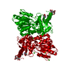
| Entry | Database: EMDB / ID: EMD-20475 | |||||||||
|---|---|---|---|---|---|---|---|---|---|---|
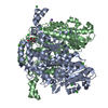
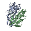
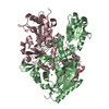
| Title | MicroED structure of proteinase K recorded on Falcon III | |||||||||
 Map data Map data | Proteinase K recorded on Falcon III | |||||||||
 Sample Sample |
| |||||||||
 Keywords Keywords | Hydrolase | |||||||||
| Function / homology |  Function and homology information Function and homology informationpeptidase K / serine-type endopeptidase activity / proteolysis / extracellular space / metal ion binding Similarity search - Function | |||||||||
| Biological species |  Parengyodontium album (fungus) Parengyodontium album (fungus) | |||||||||
| Method | electron crystallography / cryo EM / Resolution: 2.1 Å | |||||||||
 Authors Authors | Hattne J / Martynowycz MW | |||||||||
 Citation Citation | Journal: IUCrJ / Year: 2019 Title: MicroED with the Falcon III direct electron detector. Authors: Johan Hattne / Michael W Martynowycz / Pawel A Penczek / Tamir Gonen /  Abstract: Microcrystal electron diffraction (MicroED) combines crystallography and electron cryo-microscopy (cryo-EM) into a method that is applicable to high-resolution structure determination. In MicroED, ...Microcrystal electron diffraction (MicroED) combines crystallography and electron cryo-microscopy (cryo-EM) into a method that is applicable to high-resolution structure determination. In MicroED, nanosized crystals, which are often intractable using other techniques, are probed by high-energy electrons in a transmission electron microscope. Diffraction data are recorded by a camera in movie mode: the nanocrystal is continuously rotated in the beam, thus creating a sequence of frames that constitute a movie with respect to the rotation angle. Until now, diffraction-optimized cameras have mostly been used for MicroED. Here, the use of a direct electron detector that was designed for imaging is reported. It is demonstrated that data can be collected more rapidly using the Falcon III for MicroED and with markedly lower exposure than has previously been reported. The Falcon III was operated at 40 frames per second and complete data sets reaching atomic resolution were recorded in minutes. The resulting density maps to 2.1 Å resolution of the serine protease proteinase K showed no visible signs of radiation damage. It is thus demonstrated that dedicated diffraction-optimized detectors are not required for MicroED, as shown by the fact that the very same cameras that are used for imaging applications in electron microscopy, such as single-particle cryo-EM, can also be used effectively for diffraction measurements. | |||||||||
| History |
|
- Structure visualization
| Movie |
Movie viewer |
|---|---|
| Structure viewer | EM map: SurfViewMolmilJmol/JSmol |
| Supplemental images |

- Downloads & links
-EMDB archive
| Map data | emd_20475.map.gz | 1.7 MB | EMDB map data format | |
|---|---|---|---|---|
| Header (meta data) | emd-20475-v30.xmlemd-20475.xml | 14.2 KB 14.2 KB | Display Display | EMDB header |
| Images | 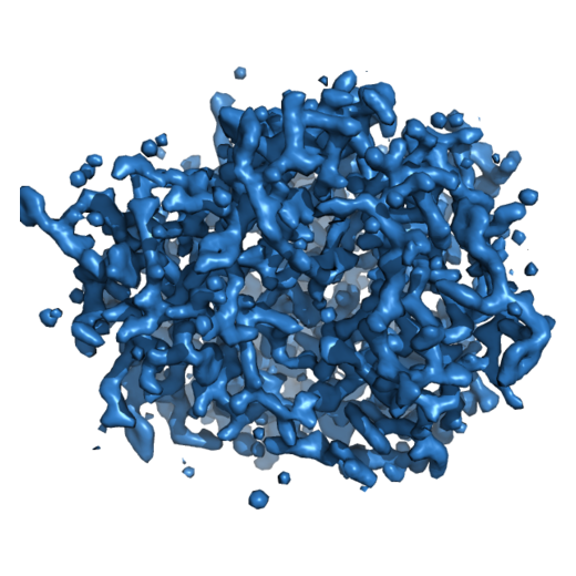 emd_20475.png emd_20475.png | 232.7 KB | ||
| Filedesc metadata | emd-20475.cif.gz | 5.4 KB | ||
| Filedesc structureFactors | emd_20475_sf.cif.gz | 382.9 KB | ||
| Archive directory |  http://ftp.pdbj.org/pub/emdb/structures/EMD-20475ftp://ftp.pdbj.org/pub/emdb/structures/EMD-20475 http://ftp.pdbj.org/pub/emdb/structures/EMD-20475ftp://ftp.pdbj.org/pub/emdb/structures/EMD-20475 | HTTPS FTP |
-Validation report
| Summary document | emd_20475_validation.pdf.gz | 610.6 KB | Display | EMDB validaton report |
|---|---|---|---|---|
| Full document | emd_20475_full_validation.pdf.gz | 610.2 KB | Display | |
| Data in XML | emd_20475_validation.xml.gz | 4.3 KB | Display | |
| Data in CIF | emd_20475_validation.cif.gz | 4.8 KB | Display | |
| Arichive directory | https://ftp.pdbj.org/pub/emdb/validation_reports/EMD-20475ftp://ftp.pdbj.org/pub/emdb/validation_reports/EMD-20475 | HTTPS FTP |
-Related structure data
| Related structure data |  6pu4MC  6pu5C C: citing same article ( M: atomic model generated by this map |
|---|---|
| Similar structure data |


-Links
| EMDB pages | EMDB (EBI/PDBe) / EMDataResource |
|---|---|
| Related items in Molecule of the Month |


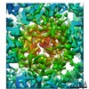
-Map
| File | Download / File: emd_20475.map.gz / Format: CCP4 / Size: 1.8 MB / Type: IMAGE STORED AS FLOATING POINT NUMBER (4 BYTES) | ||||||||||||||||||||||||||||||||||||||||||||||||||||||||||||||||||||
|---|---|---|---|---|---|---|---|---|---|---|---|---|---|---|---|---|---|---|---|---|---|---|---|---|---|---|---|---|---|---|---|---|---|---|---|---|---|---|---|---|---|---|---|---|---|---|---|---|---|---|---|---|---|---|---|---|---|---|---|---|---|---|---|---|---|---|---|---|---|
| Annotation | Proteinase K recorded on Falcon III | ||||||||||||||||||||||||||||||||||||||||||||||||||||||||||||||||||||
| Projections & slices | Image control
Images are generated by Spider. generated in cubic-lattice coordinate | ||||||||||||||||||||||||||||||||||||||||||||||||||||||||||||||||||||
| Voxel size | X: 0.70292 Å / Y: 0.70292 Å / Z: 0.7066 Å | ||||||||||||||||||||||||||||||||||||||||||||||||||||||||||||||||||||
| Density |
| ||||||||||||||||||||||||||||||||||||||||||||||||||||||||||||||||||||
| Symmetry | Space group: 96 | ||||||||||||||||||||||||||||||||||||||||||||||||||||||||||||||||||||
| Details | EMDB XML:
CCP4 map header:
| ||||||||||||||||||||||||||||||||||||||||||||||||||||||||||||||||||||
 Z (Sec.)
 X (Row.)
 Y (Col.)


-Supplemental data
- Sample components
-Entire : Proteinase K
| Entire | Name: Proteinase K |
|---|---|
| Components |
|
-Supramolecule #1: Proteinase K
| Supramolecule | Name: Proteinase K / type: complex / ID: 1 / Parent: 0 / Macromolecule list: #1 |
|---|---|
| Source (natural) | Organism: Parengyodontium album (fungus) |
| Molecular weight | Theoretical: 29.049338 KDa |
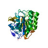
-Macromolecule #1: Proteinase K
| Macromolecule | Name: Proteinase K / type: protein_or_peptide / ID: 1 / Number of copies: 1 / Enantiomer: LEVO / EC number: peptidase K |
|---|---|
| Source (natural) | Organism: Parengyodontium album (fungus) |
| Molecular weight | Theoretical: 28.930783 KDa |
| Sequence | String: AAQTNAPWGL ARISSTSPGT STYYYDESAG QGSCVYVIDT GIEASHPEFE GRAQMVKTYY YSSRDGNGHG THCAGTVGSR TYGVAKKTQ LFGVKVLDDN GSGQYSTIIA GMDFVASDKN NRNCPKGVVA SLSLGGGYSS SVNSAAARLQ SSGVMVAVAA G NNNADARN ...String: AAQTNAPWGL ARISSTSPGT STYYYDESAG QGSCVYVIDT GIEASHPEFE GRAQMVKTYY YSSRDGNGHG THCAGTVGSR TYGVAKKTQ LFGVKVLDDN GSGQYSTIIA GMDFVASDKN NRNCPKGVVA SLSLGGGYSS SVNSAAARLQ SSGVMVAVAA G NNNADARN YSPASEPSVC TVGASDRYDR RSSFSNYGSV LDIFGPGTSI LSTWIGGSTR SISGTSMATP HVAGLAAYLM TL GKTTAAS ACRYIADTAN KGDLSNIPFG TVNLLAYNNY QA UniProtKB: Proteinase K |
-Macromolecule #2: CALCIUM ION
| Macromolecule | Name: CALCIUM ION / type: ligand / ID: 2 / Number of copies: 4 / Formula: CA |
|---|---|
| Molecular weight | Theoretical: 40.078 Da |
-Macromolecule #3: water
| Macromolecule | Name: water / type: ligand / ID: 3 / Number of copies: 92 / Formula: HOH |
|---|---|
| Molecular weight | Theoretical: 18.015 Da |
| Chemical component information |  ChemComp-HOH: |
-Experimental details
-Structure determination
| Method | cryo EM |
|---|---|
 Processing Processing | electron crystallography |
| Aggregation state | 3D array |
-Sample preparation
| Concentration | 20 mg/mL |
|---|---|
| Buffer | pH: 8 / Component - Concentration: 50.0 mM / Component - Formula: C4H11NO3 / Component - Name: Tris |
| Grid | Model: Quantifoil R2/2 / Material: COPPER / Mesh: 300 / Pretreatment - Type: GLOW DISCHARGE / Pretreatment - Time: 15 sec. |
| Vitrification | Cryogen name: ETHANE / Chamber humidity: 100 % / Chamber temperature: 298 K / Instrument: FEI VITROBOT MARK IV |
- Electron microscopy
| Microscope | FEI TALOS ARCTICA |
|---|---|
| Temperature | Min: 77.0 K / Max: 100.0 K |
| Image recording | Film or detector model: FEI FALCON III (4k x 4k) / Digitization - Dimensions - Width: 2048 pixel / Digitization - Dimensions - Height: 2048 pixel / Number grids imaged: 1 / Number real images: 387 / Number diffraction images: 387 / Average exposure time: 1.0 sec. / Average electron dose: 0.01 e/Å2 |
| Electron beam | Acceleration voltage: 200 kV / Electron source:  FIELD EMISSION GUN FIELD EMISSION GUN |
| Electron optics | Illumination mode: FLOOD BEAM / Imaging mode: DIFFRACTION / Camera length: 1700 mm |
| Sample stage | Specimen holder model: FEI TITAN KRIOS AUTOGRID HOLDER / Cooling holder cryogen: NITROGEN |
| Experimental equipment |  Model: Talos Arctica / Image courtesy: FEI Company |
+Image processing
-Atomic model buiding 1
| Initial model | PDB ID: Chain - Chain ID: A / Chain - Residue range: 106-384 / Chain - Source name: PDB / Chain - Initial model type: experimental model |
|---|---|
| Details | Electron scattering factors |
| Refinement | Space: RECIPROCAL / Protocol: OTHER / Overall B value: 13.054 |
| Output model | PDB-6pu4: |