 Movie
Movie Controller


+ Open data

- Basic information
| Entry | Database: PDB / ID: 1uaq | ||||||
|---|---|---|---|---|---|---|---|
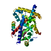
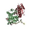
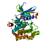
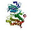
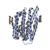
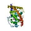
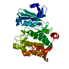
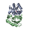
| Title | The crystal structure of yeast cytosine deaminase | ||||||
 Components Components | cytosine deaminase | ||||||
 Keywords Keywords | HYDROLASE / alpha-beta-alpha | ||||||
| Function / homology |  Function and homology information Function and homology informationcytidine metabolic process / diaminohydroxyphosphoribosylaminopyrimidine deaminase activity / cytosine deaminase / pyrimidine-containing compound salvage / : / cytosine deaminase activity / UMP salvage / cytosine metabolic process / zinc ion binding / nucleus / cytoplasm Similarity search - Function | ||||||
| Biological species |  | ||||||
| Method |  X-RAY DIFFRACTION / SYNCHROTRON / MAD / Resolution: 1.6 Å X-RAY DIFFRACTION / SYNCHROTRON / MAD / Resolution: 1.6 Å | ||||||
 Authors Authors | Ko, T.-P. / Lin, J.-J. / Hu, C.-Y. / Hsu, Y.-H. / Wang, A.H.-J. / Liaw, S.-H. | ||||||
 Citation Citation | Journal: J.Biol.Chem. / Year: 2003 Title: Crystal structure of yeast cytosine deaminase. Insights into enzyme mechanism and evolution Authors: Ko, T.-P. / Lin, J.-J. / Hu, C.-Y. / Hsu, Y.-H. / Wang, A.H.-J. / Liaw, S.-H. #1: Journal: Acta Crystallogr.,Sect.D / Year: 2003Title: Crystallization and preliminary crystallographic analysis of yeast cytosine deaminase Authors: Hsu, Y.-H. / Hu, C.-Y. / Lin, J.-J. / Liaw, S.-H. | ||||||
| History |
|
- Structure visualization
| Structure viewer | Molecule: MolmilJmol/JSmol |
|---|
- Downloads & links
-Download
| PDBx/mmCIF format | 1uaq.cif.gz | 82.4 KB | Display | PDBx/mmCIF format |
|---|---|---|---|---|
| PDB format | pdb1uaq.ent.gz | 61.4 KB | Display | PDB format |
| PDBx/mmJSON format | 1uaq.json.gz | Tree view | PDBx/mmJSON format | |
| Others |  Other downloads Other downloads |
-Validation report
| Summary document | 1uaq_validation.pdf.gz | 450.3 KB | Display | wwPDB validaton report |
|---|---|---|---|---|
| Full document | 1uaq_full_validation.pdf.gz | 453.8 KB | Display | |
| Data in XML | 1uaq_validation.xml.gz | 19.7 KB | Display | |
| Data in CIF | 1uaq_validation.cif.gz | 29.5 KB | Display | |
| Arichive directory | https://data.pdbj.org/pub/pdb/validation_reports/ua/1uaqftp://data.pdbj.org/pub/pdb/validation_reports/ua/1uaq | HTTPS FTP |
-Related structure data
| Related structure data | |
|---|---|
| Similar structure data |


-Links
 PDBj
PDBj- Assembly
| Deposited unit | 
| ||||||||
|---|---|---|---|---|---|---|---|---|---|
| 1 |
| ||||||||
| Unit cell |
|
-Components
| #1: Protein | Mass: 17530.018 Da / Num. of mol.: 2 Source method: isolated from a genetically manipulated source Source: (gene. exp.) Gene: fcy1 / Plasmid: pET-6H / Production host:  #2: Chemical |   Mass: 65.409 Da / Num. of mol.: 2 / Source method: obtained synthetically / Formula: Zn Mass: 65.409 Da / Num. of mol.: 2 / Source method: obtained synthetically / Formula: Zn#3: Chemical |   Mass: 114.103 Da / Num. of mol.: 2 / Source method: obtained synthetically / Formula: C4H6N2O2 Mass: 114.103 Da / Num. of mol.: 2 / Source method: obtained synthetically / Formula: C4H6N2O2#4: Water | ChemComp-HOH / |  Mass: 18.015 Da / Num. of mol.: 484 / Source method: isolated from a natural source / Formula: H2O Mass: 18.015 Da / Num. of mol.: 484 / Source method: isolated from a natural source / Formula: H2O |
|---|
-Experimental details
-Experiment
| Experiment | Method: X-RAY DIFFRACTION / Number of used crystals: 1 |
|---|
- Sample preparation
| Crystal | Density Matthews: 2.07 Å3/Da / Density % sol: 40.26 % | ||||||||||||||||||||||||||||||||||||
|---|---|---|---|---|---|---|---|---|---|---|---|---|---|---|---|---|---|---|---|---|---|---|---|---|---|---|---|---|---|---|---|---|---|---|---|---|---|
| Crystal grow | Temperature: 295 K / Method: vapor diffusion, hanging drop / pH: 7.5 Details: isopropanol, PEG4000, HEPES, pH 7.5, VAPOR DIFFUSION, HANGING DROP, temperature 295K | ||||||||||||||||||||||||||||||||||||
| Crystal grow | *PLUS Details: Hsu, Y.-H., (2003) Acta Crystallogr., Sect.D, 59, 950. | ||||||||||||||||||||||||||||||||||||
| Components of the solutions | *PLUS
|
-Data collection
| Diffraction | Mean temperature: 100 K |
|---|---|
| Diffraction source | Source: SYNCHROTRON / Site: SPring-8  / Beamline: BL41XU / Wavelength: 0.71 Å / Beamline: BL41XU / Wavelength: 0.71 Å |
| Detector | Type: MAR CCD 165 mm / Detector: CCD / Date: Jun 3, 2002 |
| Radiation | Monochromator: the rotated-inclined double-crystal monochromator Protocol: SINGLE WAVELENGTH / Monochromatic (M) / Laue (L): M / Scattering type: x-ray |
| Radiation wavelength | Wavelength: 0.71 Å / Relative weight: 1 |
| Reflection | Resolution: 1.6→50 Å / Num. all: 42187 / Num. obs: 42187 / % possible obs: 96.7 % / Observed criterion σ(F): 0 / Observed criterion σ(I): 0 / Redundancy: 6.6 % / Rmerge(I) obs: 0.066 / Rsym value: 0.057 / Net I/σ(I): 11.9 |
| Reflection shell | Resolution: 1.6→1.66 Å / Redundancy: 4.8 % / Rmerge(I) obs: 0.336 / Mean I/σ(I) obs: 2.9 / Num. unique all: 3945 / Rsym value: 0.297 / % possible all: 91.2 |
| Reflection | *PLUS Lowest resolution: 50 Å / Num. measured all: 277542 |
| Reflection shell | *PLUS Highest resolution: 1.6 Å / % possible obs: 91.2 % / Num. unique obs: 3945 / Num. measured obs: 18894 / Rmerge(I) obs: 0.338 |
- Processing
| Software |
| |||||||||||||||||||||||||
|---|---|---|---|---|---|---|---|---|---|---|---|---|---|---|---|---|---|---|---|---|---|---|---|---|---|---|
| Refinement | Method to determine structure: MAD / Resolution: 1.6→23.79 Å / Isotropic thermal model: isotropic / σ(F): 0 / σ(I): 0
| |||||||||||||||||||||||||
| Displacement parameters |
| |||||||||||||||||||||||||
| Refine analyze | Luzzati d res low obs: 6 Å / Luzzati sigma a obs: 0.2 Å | |||||||||||||||||||||||||
| Refinement step | Cycle: LAST / Resolution: 1.6→23.79 Å
| |||||||||||||||||||||||||
| Refine LS restraints |
| |||||||||||||||||||||||||
| LS refinement shell | Resolution: 1.6→1.66 Å /
| |||||||||||||||||||||||||
| Refinement | *PLUS Lowest resolution: 50 Å / % reflection Rfree: 5 % | |||||||||||||||||||||||||
| Solvent computation | *PLUS | |||||||||||||||||||||||||
| Displacement parameters | *PLUS | |||||||||||||||||||||||||
| LS refinement shell | *PLUS Highest resolution: 1.6 Å / Rfactor Rfree: 0.313 / Rfactor Rwork: 0.268 / Num. reflection obs: 3157 |
