 Movie
Movie Controller


+ Open data

- Basic information
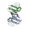
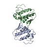
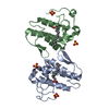
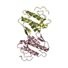
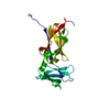
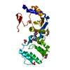
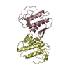
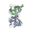
| Entry | Database: PDB / ID: 1qll | ||||||
|---|---|---|---|---|---|---|---|
| Title | Piratoxin-II (Prtx-II) - a K49 PLA2 from Bothrops pirajai | ||||||
 Components Components | PHOSPHOLIPASE A2 | ||||||
 Keywords Keywords | NEUROTOXIN / K49 PHOSPHOLIPASE A2 (PLA2) | ||||||
| Function / homology |  Function and homology information Function and homology informationA2-type glycerophospholipase activity / arachidonate secretion / lipid catabolic process / negative regulation of T cell proliferation / phospholipid metabolic process / phospholipid binding / toxin activity / calcium ion binding / extracellular region Similarity search - Function | ||||||
| Biological species |  BOTHROPS PIRAJAI (snake) BOTHROPS PIRAJAI (snake) | ||||||
| Method |  X-RAY DIFFRACTION / SYNCHROTRON / MOLECULAR REPLACEMENT / Resolution: 2.04 Å X-RAY DIFFRACTION / SYNCHROTRON / MOLECULAR REPLACEMENT / Resolution: 2.04 Å | ||||||
 Authors Authors | Lee, W.-H. / Polikarpov, I. | ||||||
 Citation Citation | Journal: Biochemistry / Year: 2001 Title: Structural Basis for Low Catalytic Activity in Lys49 Phospholipases A2-A Hypothesis: The Crystal Structure of Piratoxin II Complexed to Fatty Acid Authors: Lee, W.H. / Da Silva Giotto, M.T. / Marangoni, S. / Toyama, M.H. / Polikarpov, I. / Garratt, R.C. | ||||||
| History |
|
- Structure visualization
| Structure viewer | Molecule: MolmilJmol/JSmol |
|---|
- Downloads & links
-Download
| PDBx/mmCIF format | 1qll.cif.gz | 68.2 KB | Display | PDBx/mmCIF format |
|---|---|---|---|---|
| PDB format | pdb1qll.ent.gz | 50.8 KB | Display | PDB format |
| PDBx/mmJSON format | 1qll.json.gz | Tree view | PDBx/mmJSON format | |
| Others |  Other downloads Other downloads |
-Validation report
| Arichive directory | https://data.pdbj.org/pub/pdb/validation_reports/ql/1qllftp://data.pdbj.org/pub/pdb/validation_reports/ql/1qll | HTTPS FTP |
|---|
-Related structure data
| Related structure data |  1clpS S: Starting model for refinement |
|---|---|
| Similar structure data |

-Links
 PDBj
PDBj
- Assembly
| Deposited unit | 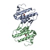
| ||||||||
|---|---|---|---|---|---|---|---|---|---|
| 1 |
| ||||||||
| Unit cell |
| ||||||||
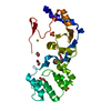
| Noncrystallographic symmetry (NCS) | NCS oper: (Code: given Matrix: (1, -0.0003, -0.0043), Vector: Details | THE ASYMMETRIC UNIT CONTAINS A HOMO-DIMERIC COMPLEX | |
-Components
| #1: Protein | Mass: 13784.216 Da / Num. of mol.: 2 / Source method: isolated from a natural source / Details: FATTY ACID TRAPPED AT THE HYDROPHOBIC CHANNEL / Source: (natural) BOTHROPS PIRAJAI (snake) / Secretion: TOTAL VENOM / References: UniProt: P82287, phospholipase A2#2: Chemical | 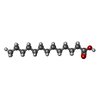  Mass: 214.344 Da / Num. of mol.: 2 / Source method: obtained synthetically / Formula: C13H26O2 Mass: 214.344 Da / Num. of mol.: 2 / Source method: obtained synthetically / Formula: C13H26O2#3: Water | ChemComp-HOH / |  Mass: 18.015 Da / Num. of mol.: 302 / Source method: isolated from a natural source / Formula: H2O Mass: 18.015 Da / Num. of mol.: 302 / Source method: isolated from a natural source / Formula: H2OCompound details | THE PHOSPHOLIPASES A2 (PLA2) CAN BE DIVIDED INTO TWO MAJOR CLASSES BASED UPON CATALYTIC ACTIVITY. ...THE PHOSPHOLIP | Has protein modification | Y | |
|---|
-Experimental details
-Experiment
| Experiment | Method: X-RAY DIFFRACTION / Number of used crystals: 2 |
|---|
- Sample preparation
| Crystal | Density Matthews: 3.17 Å3/Da / Density % sol: 61 % | ||||||||||||||||||||||||||||||
|---|---|---|---|---|---|---|---|---|---|---|---|---|---|---|---|---|---|---|---|---|---|---|---|---|---|---|---|---|---|---|---|
| Crystal grow | Temperature: 277 K / pH: 8.5 Details: 28% PEG 3350, 0.25 M LITHIUM SULFATE 0.1 M TRIS-HCL PH 8.5, AT 277 K FOR APPROX. 40 DAYS | ||||||||||||||||||||||||||||||
| Crystal grow | *PLUS Temperature: 277 K / Method: vapor diffusion, hanging drop | ||||||||||||||||||||||||||||||
| Components of the solutions | *PLUS
|
-Data collection
| Diffraction | Mean temperature: 277 K |
|---|---|
| Diffraction source | Source: SYNCHROTRON / Site: LNLS  / Beamline: D03B-MX1 / Wavelength: 1.38 / Beamline: D03B-MX1 / Wavelength: 1.38 |
| Detector | Type: MARRESEARCH / Detector: IMAGE PLATE / Date: Sep 15, 1997 |
| Radiation | Monochromator: SI(111) / Protocol: SINGLE WAVELENGTH / Monochromatic (M) / Laue (L): M / Scattering type: x-ray |
| Radiation wavelength | Wavelength: 1.38 Å / Relative weight: 1 |
| Reflection | Resolution: 2.04→20 Å / Num. obs: 6934 / % possible obs: 90.2 % / Observed criterion σ(I): 2 / Redundancy: 3.43 % / Rsym value: 0.07 / Net I/σ(I): 12.4 |
| Reflection shell | Resolution: 2.04→2.11 Å / Redundancy: 1.53 % / Mean I/σ(I) obs: 2.57 / Rsym value: 0.26 / % possible all: 72.5 |
| Reflection | *PLUS Num. measured all: 23809 / Rmerge(I) obs: 0.07 |
| Reflection shell | *PLUS % possible obs: 72.5 % / Rmerge(I) obs: 0.26 |
- Processing
| Software |
| ||||||||||||||||||||||||||||||||||||||||||||||||||||||||||||||||||||||||||||||||||||
|---|---|---|---|---|---|---|---|---|---|---|---|---|---|---|---|---|---|---|---|---|---|---|---|---|---|---|---|---|---|---|---|---|---|---|---|---|---|---|---|---|---|---|---|---|---|---|---|---|---|---|---|---|---|---|---|---|---|---|---|---|---|---|---|---|---|---|---|---|---|---|---|---|---|---|---|---|---|---|---|---|---|---|---|---|---|
| Refinement | Method to determine structure: MOLECULAR REPLACEMENT Starting model: PDB ENTRY 1CLP Resolution: 2.04→10 Å / Cross valid method: THROUGHOUT / σ(F): 2 Details: THE C-TERMINAL IS CLEARLY VISIBLE IN THE DENSITY MAP AND IT IS MAKING A DISULPHIDE BRIDGE.
| ||||||||||||||||||||||||||||||||||||||||||||||||||||||||||||||||||||||||||||||||||||
| Refinement step | Cycle: LAST / Resolution: 2.04→10 Å
| ||||||||||||||||||||||||||||||||||||||||||||||||||||||||||||||||||||||||||||||||||||
| Refine LS restraints |
| ||||||||||||||||||||||||||||||||||||||||||||||||||||||||||||||||||||||||||||||||||||
| Software | *PLUS Name: REFMAC / Classification: refinement | ||||||||||||||||||||||||||||||||||||||||||||||||||||||||||||||||||||||||||||||||||||
| Refinement | *PLUS Rfactor obs: 0.176 | ||||||||||||||||||||||||||||||||||||||||||||||||||||||||||||||||||||||||||||||||||||
| Solvent computation | *PLUS | ||||||||||||||||||||||||||||||||||||||||||||||||||||||||||||||||||||||||||||||||||||
| Displacement parameters | *PLUS |
