 Movie
Movie Controller

[English] 日本語
 Yorodumi
- PDB-1mu0: Crystal Structure of the Tricorn Interacting Factor F1 Complex wi... -
+ Open data

- Basic information
| Entry | Database: PDB / ID: 1mu0 | ||||||
|---|---|---|---|---|---|---|---|
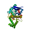
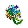
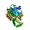
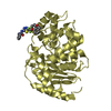
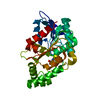
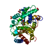
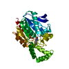
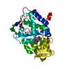
| Title | Crystal Structure of the Tricorn Interacting Factor F1 Complex with PCK | ||||||
 Components Components | Proline iminopeptidase | ||||||
 Keywords Keywords | HYDROLASE / ALPHA-BETA HYDROLASE / CAP DOMAIN / CAGED ACTIVE SITE / PROLYL PEPTIDASE | ||||||
| Function / homology |  Function and homology information Function and homology informationprolyl aminopeptidase / aminopeptidase activity / proteolysis / membrane Similarity search - Function | ||||||
| Biological species |   Thermoplasma acidophilum (acidophilic) Thermoplasma acidophilum (acidophilic) | ||||||
| Method |  X-RAY DIFFRACTION / MOLECULAR REPLACEMENT / Resolution: 2.4 Å X-RAY DIFFRACTION / MOLECULAR REPLACEMENT / Resolution: 2.4 Å | ||||||
 Authors Authors | Goettig, P. / Groll, M. / Kim, J.-S. / Huber, R. / Brandstetter, H. | ||||||
 Citation Citation | Journal: Embo J. / Year: 2002 Title: Structures of the tricorn-interacting aminopeptidase F1 with different ligands explain its catalytic mechanism Authors: Goettig, P. / Groll, M. / Kim, J.-S. / Huber, R. / Brandstetter, H. | ||||||
| History |
| ||||||
| Remark 600 | HETEROGEN THE REACTION OF PHENYLALANYLCHLOROMETHYL KETONE (PCK) WITH SER105 RESULTED IN A ... HETEROGEN THE REACTION OF PHENYLALANYLCHLOROMETHYL KETONE (PCK) WITH SER105 RESULTED IN A COVALENTLY BOUND HETEROMOLECULE (PHK). |
- Structure visualization
| Structure viewer | Molecule: MolmilJmol/JSmol |
|---|
- Downloads & links
-Download
| PDBx/mmCIF format | 1mu0.cif.gz | 71.6 KB | Display | PDBx/mmCIF format |
|---|---|---|---|---|
| PDB format | pdb1mu0.ent.gz | 52.7 KB | Display | PDB format |
| PDBx/mmJSON format | 1mu0.json.gz | Tree view | PDBx/mmJSON format | |
| Others |  Other downloads Other downloads |
-Validation report
| Arichive directory | https://data.pdbj.org/pub/pdb/validation_reports/mu/1mu0ftp://data.pdbj.org/pub/pdb/validation_reports/mu/1mu0 | HTTPS FTP |
|---|
-Related structure data
| Related structure data | 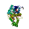 1mt3SC 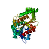 1mtzC S: Starting model for refinement C: citing same article ( |
|---|---|
| Similar structure data |


-Links
 PDBj
PDBj


- Assembly
| Deposited unit | 
| ||||||||
|---|---|---|---|---|---|---|---|---|---|
| 1 |
| ||||||||
| Unit cell |
| ||||||||
| Details | The biological assembly is the monomer in the asymmetric unit |
-Components
| #1: Protein | Mass: 33530.090 Da / Num. of mol.: 1 Source method: isolated from a genetically manipulated source Source: (gene. exp.) Thermoplasma acidophilum (acidophilic) / Gene: TA0830 / Plasmid: pRset6c / Production host:  |
|---|---|
| #2: Chemical | ChemComp-PHK / (  Mass: 199.677 Da / Num. of mol.: 1 / Source method: obtained synthetically / Formula: C10H14ClNO Mass: 199.677 Da / Num. of mol.: 1 / Source method: obtained synthetically / Formula: C10H14ClNO |
| #3: Water | ChemComp-HOH /  Mass: 18.015 Da / Num. of mol.: 37 / Source method: isolated from a natural source / Formula: H2O Mass: 18.015 Da / Num. of mol.: 37 / Source method: isolated from a natural source / Formula: H2O |
| Has protein modification | Y |
-Experimental details
-Experiment
| Experiment | Method: X-RAY DIFFRACTION / Number of used crystals: 1 |
|---|
- Sample preparation
| Crystal | Density Matthews: 2.13 Å3/Da / Density % sol: 42.27 % | ||||||||||||||||||||||||||||||||||||||||||
|---|---|---|---|---|---|---|---|---|---|---|---|---|---|---|---|---|---|---|---|---|---|---|---|---|---|---|---|---|---|---|---|---|---|---|---|---|---|---|---|---|---|---|---|
| Crystal grow | Temperature: 293 K / Method: vapor diffusion, hanging drop / pH: 6 Details: 12% PEG 6000, 100 mM Bis-Tris-HCl, pH 6.0, VAPOR DIFFUSION, HANGING DROP, temperature 293K | ||||||||||||||||||||||||||||||||||||||||||
| Crystal grow | *PLUS Method: vapor diffusion | ||||||||||||||||||||||||||||||||||||||||||
| Components of the solutions | *PLUS
|
-Data collection
| Diffraction | Mean temperature: 100 K |
|---|---|
| Diffraction source | Source: ROTATING ANODE / Type: RIGAKU RU300 / Wavelength: 1.5418 Å |
| Detector | Type: MARRESEARCH / Detector: IMAGE PLATE / Date: Jan 10, 2002 / Details: Osmic mirrors |
| Radiation | Protocol: SINGLE WAVELENGTH / Monochromatic (M) / Laue (L): M / Scattering type: x-ray |
| Radiation wavelength | Wavelength: 1.5418 Å / Relative weight: 1 |
| Reflection | Resolution: 2.2→20 Å / Num. all: 12507 / Num. obs: 12507 / % possible obs: 85.7 % / Observed criterion σ(F): 1 / Observed criterion σ(I): 2 / Biso Wilson estimate: 22.1 Å2 / Net I/σ(I): 3.2 |
| Reflection | *PLUS Highest resolution: 2.4 Å / Lowest resolution: 20 Å / Num. obs: 11344 |
- Processing
| Software |
| |||||||||||||||||||||||||
|---|---|---|---|---|---|---|---|---|---|---|---|---|---|---|---|---|---|---|---|---|---|---|---|---|---|---|
| Refinement | Method to determine structure: MOLECULAR REPLACEMENT Starting model: PDB ENTRY 1MT3 Resolution: 2.4→20 Å / Cross valid method: THROUGHOUT / σ(F): 0 / Stereochemistry target values: Engh & Huber
| |||||||||||||||||||||||||
| Displacement parameters | Biso mean: 23.5 Å2
| |||||||||||||||||||||||||
| Refinement step | Cycle: LAST / Resolution: 2.4→20 Å
| |||||||||||||||||||||||||
| Refine LS restraints |
| |||||||||||||||||||||||||
| Refinement | *PLUS Num. reflection obs: 9958 | |||||||||||||||||||||||||
| Solvent computation | *PLUS | |||||||||||||||||||||||||
| Displacement parameters | *PLUS | |||||||||||||||||||||||||
| Refine LS restraints | *PLUS Type: c_angle_deg / Dev ideal: 0.948 |
