 Movie
Movie Controller

[English] 日本語
 Yorodumi
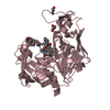
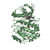
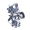
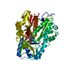
- PDB-1m4g: Aminoglycoside 2'-N-acetyltransferase from Mycobacterium tubercul... -
+ Open data

- Basic information
| Entry | Database: PDB / ID: 1m4g | ||||||
|---|---|---|---|---|---|---|---|
| Title | Aminoglycoside 2'-N-acetyltransferase from Mycobacterium tuberculosis-Complex with Coenzyme A and Ribostamycin | ||||||
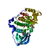
 Components Components | Aminoglycoside 2'-N-acetyltransferase | ||||||
 Keywords Keywords | TRANSFERASE / COA BINDING MOTIF | ||||||
| Function / homology |  Function and homology information Function and homology informationaminoglycoside 2'-N-acetyltransferase activity / aminoglycoside antibiotic catabolic process / aminoglycoside N-acetyltransferase activity / N-acetyltransferase activity / Transferases; Acyltransferases; Transferring groups other than aminoacyl groups / response to antibiotic / plasma membrane Similarity search - Function | ||||||
| Biological species |   Mycobacterium tuberculosis (bacteria) Mycobacterium tuberculosis (bacteria) | ||||||
| Method |  X-RAY DIFFRACTION / MOLECULAR REPLACEMENT / Resolution: 1.8 Å X-RAY DIFFRACTION / MOLECULAR REPLACEMENT / Resolution: 1.8 Å | ||||||
 Authors Authors | Vetting, M.W. / Hegde, S.S. / Javid-Majd, F. / Blanchard, J.S. / Roderick, S.L. | ||||||
 Citation Citation | Journal: Nat.Struct.Biol. / Year: 2002 Title: Aminoglycoside 2'-N-acetyltransferase from Mycobacterium tuberculosis in complex with coenzyme A and aminoglycoside substrates. Authors: Vetting, M.W. / Hegde, S.S. / Javid-Majd, F. / Blanchard, J.S. / Roderick, S.L. #1: Journal: J.Biol.Chem. / Year: 2001Title: Overexpression and Mechanistic Analysis of Chromosomally Encoded Aminoglycoside 2'-N-Acetyltransferase (AAC(2')-Ic) from Mycobacterium tuberculosis Authors: Hegde, S.S. / Javid-Majd, F. / Blanchard, J.S. | ||||||
| History |
|
- Structure visualization
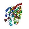
| Structure viewer | Molecule: MolmilJmol/JSmol |
|---|
- Downloads & links
-Download
| PDBx/mmCIF format | 1m4g.cif.gz | 95.9 KB | Display | PDBx/mmCIF format |
|---|---|---|---|---|
| PDB format | pdb1m4g.ent.gz | 72.7 KB | Display | PDB format |
| PDBx/mmJSON format | 1m4g.json.gz | Tree view | PDBx/mmJSON format | |
| Others |  Other downloads Other downloads |
-Validation report
| Arichive directory | https://data.pdbj.org/pub/pdb/validation_reports/m4/1m4gftp://data.pdbj.org/pub/pdb/validation_reports/m4/1m4g | HTTPS FTP |
|---|
-Related structure data
| Related structure data |  1m44SC  1m4dC 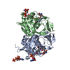 1m4iC S: Starting model for refinement C: citing same article ( |
|---|---|
| Similar structure data |


-Links
 PDBj
PDBj


- Assembly
| Deposited unit | 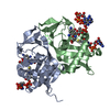
| ||||||||
|---|---|---|---|---|---|---|---|---|---|
| 1 |
| ||||||||
| Unit cell |
| ||||||||
| Details | The biological assembly is a dimer as contained in the assymetric unit |
-Components
| #1: Protein | Mass: 20063.699 Da / Num. of mol.: 2 Source method: isolated from a genetically manipulated source Source: (gene. exp.) Mycobacterium tuberculosis (bacteria) / Strain: H37Rv / Production host: References: UniProt: P0A5N0, UniProt: P9WQG9*PLUS, Transferases; Acyltransferases; Transferring groups other than aminoacyl groups #2: Chemical |   Mass: 767.534 Da / Num. of mol.: 2 / Source method: obtained synthetically / Formula: C21H36N7O16P3S Mass: 767.534 Da / Num. of mol.: 2 / Source method: obtained synthetically / Formula: C21H36N7O16P3S#3: Chemical | 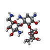  Mass: 454.473 Da / Num. of mol.: 2 / Source method: obtained synthetically / Formula: C17H34N4O10 / Comment: antibiotic*YM Mass: 454.473 Da / Num. of mol.: 2 / Source method: obtained synthetically / Formula: C17H34N4O10 / Comment: antibiotic*YM#4: Chemical |   Mass: 507.181 Da / Num. of mol.: 2 / Source method: obtained synthetically / Formula: C10H16N5O13P3 Mass: 507.181 Da / Num. of mol.: 2 / Source method: obtained synthetically / Formula: C10H16N5O13P3#5: Water | ChemComp-HOH / |  Mass: 18.015 Da / Num. of mol.: 311 / Source method: isolated from a natural source / Formula: H2O Mass: 18.015 Da / Num. of mol.: 311 / Source method: isolated from a natural source / Formula: H2O |
|---|
-Experimental details
-Experiment
| Experiment | Method: X-RAY DIFFRACTION / Number of used crystals: 1 |
|---|
- Sample preparation
| Crystal | Density Matthews: 2.58 Å3/Da / Density % sol: 52.3 % | ||||||||||||||||||||||||||||||
|---|---|---|---|---|---|---|---|---|---|---|---|---|---|---|---|---|---|---|---|---|---|---|---|---|---|---|---|---|---|---|---|
| Crystal grow | Temperature: 298 K / Method: vapor diffusion, hanging drop / pH: 6.5 Details: Ammonium Sulfate, ADA, Coenzyme A, Ribostamycin, pH 6.5, VAPOR DIFFUSION, HANGING DROP, temperature 298K | ||||||||||||||||||||||||||||||
| Crystal grow | *PLUS Method: unknown | ||||||||||||||||||||||||||||||
| Components of the solutions | *PLUS
|
-Data collection
| Diffraction | Mean temperature: 298 K |
|---|---|
| Diffraction source | Source: ROTATING ANODE / Type: RIGAKU RU300 / Wavelength: 1.54 Å |
| Detector | Type: RIGAKU RAXIS IV / Detector: IMAGE PLATE / Date: Feb 2, 2002 / Details: Confocal Optics |
| Radiation | Protocol: SINGLE WAVELENGTH / Monochromatic (M) / Laue (L): M / Scattering type: x-ray |
| Radiation wavelength | Wavelength: 1.54 Å / Relative weight: 1 |
| Reflection | Resolution: 1.8→24.09 Å / Num. all: 39240 / Num. obs: 38456 / % possible obs: 98 % / Observed criterion σ(I): -3 / Redundancy: 3.5 % / Biso Wilson estimate: 14.5 Å2 / Rsym value: 0.05 / Net I/σ(I): 16.5 |
| Reflection shell | Resolution: 1.8→1.88 Å / Redundancy: 3.1 % / Mean I/σ(I) obs: 8.7 / Num. unique all: 4121 / Rsym value: 0.132 / % possible all: 96.3 |
| Reflection | *PLUS Lowest resolution: 50 Å / Num. obs: 38407 / % possible obs: 98.1 % / Rmerge(I) obs: 0.05 |
| Reflection shell | *PLUS % possible obs: 96.3 % / Rmerge(I) obs: 0.132 |
- Processing
| Software |
| |||||||||||||||||||||||||
|---|---|---|---|---|---|---|---|---|---|---|---|---|---|---|---|---|---|---|---|---|---|---|---|---|---|---|
| Refinement | Method to determine structure: MOLECULAR REPLACEMENT Starting model: PDB ID 1M44 Resolution: 1.8→50 Å / Cross valid method: THROUGHOUT / σ(F): 0 / Stereochemistry target values: Engh & Huber
| |||||||||||||||||||||||||
| Refine analyze | Luzzati coordinate error obs: 0.159 Å | |||||||||||||||||||||||||
| Refinement step | Cycle: LAST / Resolution: 1.8→50 Å
| |||||||||||||||||||||||||
| Refine LS restraints |
| |||||||||||||||||||||||||
| LS refinement shell | Resolution: 1.8→1.88 Å
| |||||||||||||||||||||||||
| Refinement | *PLUS Lowest resolution: 50 Å / % reflection Rfree: 5 % / Rfactor Rfree: 0.202 / Rfactor Rwork: 0.165 | |||||||||||||||||||||||||
| Solvent computation | *PLUS | |||||||||||||||||||||||||
| Displacement parameters | *PLUS | |||||||||||||||||||||||||
| Refine LS restraints | *PLUS Type: c_angle_deg / Dev ideal: 2.1 | |||||||||||||||||||||||||
| LS refinement shell | *PLUS Rfactor Rfree: 0.23 / Rfactor Rwork: 0.196 |
