 Movie
Movie Controller

[English] 日本語
 Yorodumi
- PDB-1lv8: Crystal structure of calf spleen purine nucleoside phosphorylase ... -
+ Open data

- Basic information
| Entry | Database: PDB / ID: 1lv8 | ||||||
|---|---|---|---|---|---|---|---|
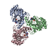
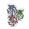
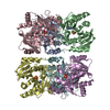
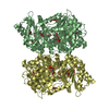
| Title | Crystal structure of calf spleen purine nucleoside phosphorylase in a new space group with full trimer in the asymmetric unit | ||||||
 Components Components | PURINE NUCLEOSIDE PHOSPHORYLASE | ||||||
 Keywords Keywords | TRANSFERASE / PNP / PURIE NUCLEOSIDE PHOSPHORYLASE / PENTOSYLTRANSFERASE / NE 2 / 6-DIAMINOPURINE MULTISUBSTRATE ANALOGUE INHIBITOR | ||||||
| Function / homology |  Function and homology information Function and homology informationguanosine phosphorylase activity / purine-nucleoside phosphorylase activity / purine-nucleoside phosphorylase / purine ribonucleoside salvage / cytosol / cytoplasm Similarity search - Function | ||||||
| Biological species |  | ||||||
| Method |  X-RAY DIFFRACTION / SYNCHROTRON / MOLECULAR REPLACEMENT / Resolution: 2.3 Å X-RAY DIFFRACTION / SYNCHROTRON / MOLECULAR REPLACEMENT / Resolution: 2.3 Å | ||||||
 Authors Authors | Bzowska, A. / Koellner, G. / Wielgus-Kutrowska, B. / Stroh, A. / Raszewski, G. / Holy, A. / Steiner, T. / Frank, J. | ||||||
 Citation Citation | Journal: J.Mol.Biol. / Year: 2004 Title: Crystal structure of calf spleen purine nucleoside phosphorylase with two full trimers in the asymmetric unit: important implications for the mechanism of catalysis Authors: Bzowska, A. / Koellner, G. / Wielgus-Kutrowska, B. / Stroh, A. / Raszewski, G. / Holy, A. / Steiner, T. / Frank, J. #1: Journal: NUCLEOSIDES NUCLEOTIDES NUCLEIC ACIDS / Year: 2003Title: Crystal Structure of Calf Spleen Purine Nucleoside in a Complex with Multisubstrate Analogue Inhibitor with 2,6-Diaminopurine Aglycone Authors: Koellner, G. / Stroh, A. / Raszewski, G. / Holy, A. / Bzowska, A. #2: Journal: J.Mol.Biol. / Year: 1997Title: Crystals Structure of Calf Spleen Purine Nucleoside Phosphorylase in a Complex with Hypoxanthine at 2.15 A Resolution Authors: Koellner, G. / Luic, M. / Shugar, D. / Saenger, W. / Bzowska, A. #3: Journal: ACTA CRYSTALLOGR.,SECT.D / Year: 2001Title: Calf Spleen Purine Nucleoside Phsophorylase: Crystal Structure of its Ternary Complex with an N(7)-Acycloguanosine Inhibitor and a Phosphate Anion Authors: Luic, M. / Koellner, G. / Shugar, D. / Saenger, W. / Bzowska, A. | ||||||
| History |
|
- Structure visualization
| Structure viewer | Molecule: MolmilJmol/JSmol |
|---|
- Downloads & links
-Download
| PDBx/mmCIF format | 1lv8.cif.gz | 344.8 KB | Display | PDBx/mmCIF format |
|---|---|---|---|---|
| PDB format | pdb1lv8.ent.gz | 276.7 KB | Display | PDB format |
| PDBx/mmJSON format | 1lv8.json.gz | Tree view | PDBx/mmJSON format | |
| Others |  Other downloads Other downloads |
-Validation report
| Arichive directory | https://data.pdbj.org/pub/pdb/validation_reports/lv/1lv8ftp://data.pdbj.org/pub/pdb/validation_reports/lv/1lv8 | HTTPS FTP |
|---|
-Related structure data
| Related structure data | 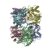 1lvuC 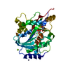 1fxuS S: Starting model for refinement C: citing same article ( |
|---|---|
| Similar structure data |


-Links
 PDBj
PDBj- Assembly
| Deposited unit | 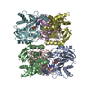
| ||||||||
|---|---|---|---|---|---|---|---|---|---|
| 1 |
| ||||||||
| Unit cell |
|
-Components
| #1: Protein | Mass: 32126.557 Da / Num. of mol.: 6 / Source method: isolated from a natural source / Source: (natural) References: UniProt: P55859, purine-nucleoside phosphorylase #2: Chemical | ChemComp-CA /   Mass: 40.078 Da / Num. of mol.: 6 / Source method: obtained synthetically / Formula: Ca Mass: 40.078 Da / Num. of mol.: 6 / Source method: obtained synthetically / Formula: Ca#3: Chemical | ChemComp-9PP /   Mass: 300.211 Da / Num. of mol.: 6 / Source method: obtained synthetically / Formula: C9H13N6O4P Mass: 300.211 Da / Num. of mol.: 6 / Source method: obtained synthetically / Formula: C9H13N6O4P#4: Water | ChemComp-HOH / |  Mass: 18.015 Da / Num. of mol.: 724 / Source method: isolated from a natural source / Formula: H2O Mass: 18.015 Da / Num. of mol.: 724 / Source method: isolated from a natural source / Formula: H2O |
|---|
-Experimental details
-Experiment
| Experiment | Method: X-RAY DIFFRACTION / Number of used crystals: 1 |
|---|
- Sample preparation
| Crystal | Density Matthews: 2.44 Å3/Da / Density % sol: 49.61 % | |||||||||||||||||||||||||||||||||||
|---|---|---|---|---|---|---|---|---|---|---|---|---|---|---|---|---|---|---|---|---|---|---|---|---|---|---|---|---|---|---|---|---|---|---|---|---|
| Crystal grow | Temperature: 298 K / Method: vapor diffusion, hanging drop / pH: 7.25 Details: HEPES, PEG400, CaCl2, pH 7.25, VAPOR DIFFUSION, HANGING DROP, temperature 298K | |||||||||||||||||||||||||||||||||||
| Crystal grow | *PLUS Temperature: 18 ℃ / Method: vapor diffusion, hanging drop | |||||||||||||||||||||||||||||||||||
| Components of the solutions | *PLUS
|
-Data collection
| Diffraction | Mean temperature: 100 K |
|---|---|
| Diffraction source | Source: SYNCHROTRON / Site: EMBL/DESY, HAMBURG  / Beamline: X11 / Wavelength: 0.9101 Å / Beamline: X11 / Wavelength: 0.9101 Å |
| Detector | Type: MARRESEARCH / Detector: IMAGE PLATE / Date: Sep 1, 2000 |
| Radiation | Protocol: SINGLE WAVELENGTH / Monochromatic (M) / Laue (L): M / Scattering type: x-ray |
| Radiation wavelength | Wavelength: 0.9101 Å / Relative weight: 1 |
| Reflection | Resolution: 2.3→19.9 Å / Num. obs: 98387 / % possible obs: 99.7 % / Observed criterion σ(I): 15.7 / Biso Wilson estimate: 32.8 Å2 / Rmerge(I) obs: 0.055 |
| Reflection shell | Resolution: 2.3→2.44 Å / Num. unique all: 98387 / % possible all: 99.7 |
| Reflection | *PLUS Lowest resolution: 20 Å / Num. obs: 84155 |
| Reflection shell | *PLUS % possible obs: 99.7 % / Rmerge(I) obs: 0.377 |
- Processing
| Software |
| ||||||||||||||||||||||||||||||||||||
|---|---|---|---|---|---|---|---|---|---|---|---|---|---|---|---|---|---|---|---|---|---|---|---|---|---|---|---|---|---|---|---|---|---|---|---|---|---|
| Refinement | Method to determine structure: MOLECULAR REPLACEMENT Starting model: 1FXU Resolution: 2.3→19.85 Å / Rfactor Rfree error: 0.003 / Isotropic thermal model: RESTRAINED / Cross valid method: THROUGHOUT / σ(F): 1 / Stereochemistry target values: Engh & Huber Details: PROTEIN WAS CO-CRYSTALLIZED WITH 9PP FROM 19-31% PEG-400 IN 100 MM HEPES BUFFER, PH 7.25; 100MM CACL2 PROTEIN WAS MIXED WITH 9PP AT A 1:5 (PNP TRIMER : INHIBITOR) MOLAR RATIO
| ||||||||||||||||||||||||||||||||||||
| Solvent computation | Solvent model: FLAT MODEL / Bsol: 44.0256 Å2 / ksol: 0.323506 e/Å3 | ||||||||||||||||||||||||||||||||||||
| Displacement parameters | Biso mean: 43 Å2
| ||||||||||||||||||||||||||||||||||||
| Refine analyze | Luzzati coordinate error free: 0.36 Å / Luzzati sigma a free: 0.35 Å | ||||||||||||||||||||||||||||||||||||
| Refinement step | Cycle: LAST / Resolution: 2.3→19.85 Å
| ||||||||||||||||||||||||||||||||||||
| Refine LS restraints |
| ||||||||||||||||||||||||||||||||||||
| LS refinement shell | Resolution: 2.3→2.44 Å / Rfactor Rfree error: 0.01 / Total num. of bins used: 6
| ||||||||||||||||||||||||||||||||||||
| Xplor file |
| ||||||||||||||||||||||||||||||||||||
| Refinement | *PLUS Highest resolution: 2.3 Å / Lowest resolution: 20 Å / % reflection Rfree: 10.3 % | ||||||||||||||||||||||||||||||||||||
| Solvent computation | *PLUS | ||||||||||||||||||||||||||||||||||||
| Displacement parameters | *PLUS | ||||||||||||||||||||||||||||||||||||
| Refine LS restraints | *PLUS
|
