 Movie
Movie Controller

[English] 日本語
 Yorodumi
- PDB-1yqu: Escherichia coli purine nucleoside phosphorylase II, the product ... -
+ Open data

- Basic information
| Entry | Database: PDB / ID: 1yqu | ||||||
|---|---|---|---|---|---|---|---|
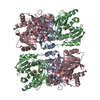
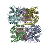
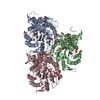
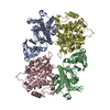
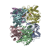
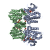
| Title | Escherichia coli purine nucleoside phosphorylase II, the product of the xapA gene | ||||||
 Components Components | Xanthosine phosphorylase | ||||||
 Keywords Keywords | TRANSFERASE / purine nucleoside phosphorylase guanine xanthine | ||||||
| Function / homology |  Function and homology information Function and homology informationguanosine catabolic process / xanthosine catabolic process / inosine nucleosidase activity / deoxyguanosine catabolic process / deoxyinosine catabolic process / inosine catabolic process / nucleobase-containing small molecule metabolic process / NAD+ biosynthetic process via the salvage pathway / guanosine phosphorylase activity / purine-nucleoside phosphorylase ...guanosine catabolic process / xanthosine catabolic process / inosine nucleosidase activity / deoxyguanosine catabolic process / deoxyinosine catabolic process / inosine catabolic process / nucleobase-containing small molecule metabolic process / NAD+ biosynthetic process via the salvage pathway / guanosine phosphorylase activity / purine-nucleoside phosphorylase / nucleobase-containing small molecule interconversion / purine-nucleoside phosphorylase activity / purine nucleoside catabolic process / protein hexamerization / identical protein binding / cytoplasm Similarity search - Function | ||||||
| Biological species |  | ||||||
| Method |  X-RAY DIFFRACTION / SYNCHROTRON / MOLECULAR REPLACEMENT / Resolution: 3.1 Å X-RAY DIFFRACTION / SYNCHROTRON / MOLECULAR REPLACEMENT / Resolution: 3.1 Å | ||||||
 Authors Authors | Dandanell, G. / Szczepanowski, R.H. / Kierdaszuk, B. / Shugar, D. / Bochtler, M. | ||||||
 Citation Citation | Journal: J.Mol.Biol. / Year: 2005 Title: Escherichia coli purine nucleoside phosphorylase II, the product of the xapA gene Authors: Dandanell, G. / Szczepanowski, R.H. / Kierdaszuk, B. / Shugar, D. / Bochtler, M. | ||||||
| History |
|
- Structure visualization
| Structure viewer | Molecule: MolmilJmol/JSmol |
|---|
- Downloads & links
-Download
| PDBx/mmCIF format | 1yqu.cif.gz | 146.1 KB | Display | PDBx/mmCIF format |
|---|---|---|---|---|
| PDB format | pdb1yqu.ent.gz | 117 KB | Display | PDB format |
| PDBx/mmJSON format | 1yqu.json.gz | Tree view | PDBx/mmJSON format | |
| Others |  Other downloads Other downloads |
-Validation report
| Arichive directory | https://data.pdbj.org/pub/pdb/validation_reports/yq/1yquftp://data.pdbj.org/pub/pdb/validation_reports/yq/1yqu | HTTPS FTP |
|---|
-Related structure data
| Related structure data |  1yqqC 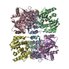 1yr3C 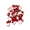 4pnpS C: citing same article ( S: Starting model for refinement |
|---|---|
| Similar structure data |


-Links
 PDBj
PDBj

- Assembly
| Deposited unit | 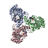
| ||||||||
|---|---|---|---|---|---|---|---|---|---|
| 1 | 
| ||||||||
| Unit cell |
| ||||||||
| Details | a hexamer with 32 point symmetry that results from the dimerization of trimers |
-Components
| #1: Protein | Mass: 29865.590 Da / Num. of mol.: 3 Source method: isolated from a genetically manipulated source Source: (gene. exp.) References: UniProt: P45563, Transferases; Glycosyltransferases; Pentosyltransferases #2: Chemical |   Mass: 94.971 Da / Num. of mol.: 3 / Source method: obtained synthetically / Formula: PO4 Mass: 94.971 Da / Num. of mol.: 3 / Source method: obtained synthetically / Formula: PO4#3: Chemical |   Mass: 151.126 Da / Num. of mol.: 3 / Source method: obtained synthetically / Formula: C5H5N5O Mass: 151.126 Da / Num. of mol.: 3 / Source method: obtained synthetically / Formula: C5H5N5O |
|---|
-Experimental details
-Experiment
| Experiment | Method: X-RAY DIFFRACTION / Number of used crystals: 1 |
|---|
- Sample preparation
| Crystal | Density Matthews: 2.1 Å3/Da / Density % sol: 41 % |
|---|---|
| Crystal grow | Temperature: 294 K / Method: vapor diffusion, sitting drop / pH: 8.2 Details: Tris, PEG4K, pH 8.2, VAPOR DIFFUSION, SITTING DROP, temperature 294K |
-Data collection
| Diffraction source | Source: SYNCHROTRON / Site: EMBL/DESY, HAMBURG  / Beamline: X11 / Wavelength: 0.811 Å / Beamline: X11 / Wavelength: 0.811 Å |
|---|---|
| Detector | Type: MARRESEARCH / Detector: CCD / Date: Mar 4, 2002 |
| Radiation | Monochromator: triangular monochromator, bent mirror (information from the Web-site) Protocol: SINGLE WAVELENGTH / Monochromatic (M) / Laue (L): M / Scattering type: x-ray |
| Radiation wavelength | Wavelength: 0.811 Å / Relative weight: 1 |
| Reflection | Resolution: 3.1→25 Å / Num. all: 15055 / Num. obs: 15055 / % possible obs: 99.9 % / Observed criterion σ(F): 0 / Observed criterion σ(I): 0 / Redundancy: 7 % / Biso Wilson estimate: 64 Å2 / Rmerge(I) obs: 0.126 / Rsym value: 0.126 / Net I/σ(I): 11 |
| Reflection shell | Resolution: 3.1→3.21 Å / Redundancy: 6 % / Rmerge(I) obs: 0.382 / Mean I/σ(I) obs: 2.8 / Num. unique all: 1436 / Rsym value: 0.382 / % possible all: 100 |
- Processing
| Software |
| ||||||||||||||||||||
|---|---|---|---|---|---|---|---|---|---|---|---|---|---|---|---|---|---|---|---|---|---|
| Refinement | Method to determine structure: MOLECULAR REPLACEMENT Starting model: pdb entry 4PNP Resolution: 3.1→25 Å / Isotropic thermal model: Isotropic / Cross valid method: THROUGHOUT / σ(F): 0 / σ(I): 0 / Stereochemistry target values: Engh & Huber
| ||||||||||||||||||||
| Displacement parameters | Biso mean: 42 Å2
| ||||||||||||||||||||
| Refinement step | Cycle: LAST / Resolution: 3.1→25 Å
|
