+ Open data
- Basic information
| Entry | Database: PDB / ID: 1kyi | ||||||
|---|---|---|---|---|---|---|---|
| Title | HslUV (H. influenzae)-NLVS Vinyl Sulfone Inhibitor Complex | ||||||
 Components Components |
| ||||||
 Keywords Keywords | CHAPERONE/HYDROLASE / prokaryotic proteasome / protease / AAA-protein / ATP-dependent chaperone / Clp/Hsp100 / vinyl sulfone inhibitor / CHAPERONE-HYDROLASE COMPLEX | ||||||
| Function / homology |  Function and homology information Function and homology informationHslU-HslV peptidase / HslUV protease complex / proteasome-activating activity / proteasome core complex / protein unfolding / threonine-type endopeptidase activity / : / peptidase activity / ATP hydrolysis activity / ATP binding ...HslU-HslV peptidase / HslUV protease complex / proteasome-activating activity / proteasome core complex / protein unfolding / threonine-type endopeptidase activity / : / peptidase activity / ATP hydrolysis activity / ATP binding / metal ion binding / cytoplasm Similarity search - Function | ||||||
| Biological species |  Haemophilus influenzae (bacteria) Haemophilus influenzae (bacteria) | ||||||
| Method |  X-RAY DIFFRACTION / X-RAY DIFFRACTION /  SYNCHROTRON / SYNCHROTRON /  MOLECULAR REPLACEMENT / Resolution: 3.1 Å MOLECULAR REPLACEMENT / Resolution: 3.1 Å | ||||||
 Authors Authors | Sousa, M.C. / Kessler, B.M. / Overkleeft, H.S. / McKay, D.B. | ||||||
 Citation Citation |  Journal: J.Mol.Biol. / Year: 2002 Journal: J.Mol.Biol. / Year: 2002Title: Crystal Structure of HslUV Complexed with a Vinyl Sulfone Inhibitor: Corroboration of a Proposed Mechanism of Allosteric Activation of HslV by HslU Authors: Sousa, M.C. / Kessler, B.M. / Overkleeft, H.S. / McKay, D.B. #1:  Journal: Cell(Cambridge,Mass.) / Year: 2000 Journal: Cell(Cambridge,Mass.) / Year: 2000Title: Crystal and Solution Structures of an HslUV Protease-Chaperone Complex Authors: Sousa, M.C. / Trame, C.B. / Tsuruta, H. / Wilbanks, S.M. / Reddy, V.S. / McKay, D.B. #2:  Journal: Acta Crystallogr.,Sect.D / Year: 2001 Journal: Acta Crystallogr.,Sect.D / Year: 2001Title: Structure of Haemophilus influenzae HslV protein at 1.9 A resolution, revealing a cation-binding site near the catalytic site Authors: Sousa, M.C. / McKay, D.B. #3:  Journal: Acta Crystallogr.,Sect.D / Year: 2001 Journal: Acta Crystallogr.,Sect.D / Year: 2001Title: Structure of Haemophilus influenzae HslU protein in crystals with one-dimensional disorder twinning Authors: Trame, C.B. / McKay, D.B. | ||||||
| History |
| ||||||
| Remark 600 | HETEROGEN ATP 450 IS ASSOCIATED WITH CHAIN A. ATP 451 IS ASSOCIATED WITH CHAIN B. ATP 452 IS ...HETEROGEN ATP 450 IS ASSOCIATED WITH CHAIN A. ATP 451 IS ASSOCIATED WITH CHAIN B. ATP 452 IS ASSOCIATED WITH CHAIN C. ATP 453 IS ASSOCIATED WITH CHAIN D. ATP 454 IS ASSOCIATED WITH CHAIN E. ATP 455 IS ASSOCIATED WITH CHAIN F. ATP 456 IS ASSOCIATED WITH CHAIN S. ATP 457 IS ASSOCIATED WITH CHAIN T. ATP 458 IS ASSOCIATED WITH CHAIN U. ATP 459 IS ASSOCIATED WITH CHAIN V. ATP 460 IS ASSOCIATED WITH CHAIN W. ATP 461 IS ASSOCIATED WITH CHAIN X. |
- Structure visualization
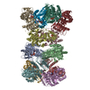
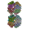
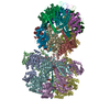
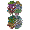
| Structure viewer | Molecule:  Molmil Molmil Jmol/JSmol Jmol/JSmol |
|---|
- Downloads & links
- Download
| PDBx/mmCIF format |  1kyi.cif.gz 1kyi.cif.gz | 1.1 MB | Display |  PDBx/mmCIF format PDBx/mmCIF format |
|---|---|---|---|---|
| PDB format |  pdb1kyi.ent.gz pdb1kyi.ent.gz | 927.3 KB | Display |  PDB format PDB format |
| PDBx/mmJSON format |  1kyi.json.gz 1kyi.json.gz | Tree view |  PDBx/mmJSON format PDBx/mmJSON format | |
| Others |  Other downloads Other downloads |
-Validation report
| Arichive directory |  https://data.pdbj.org/pub/pdb/validation_reports/ky/1kyi https://data.pdbj.org/pub/pdb/validation_reports/ky/1kyi ftp://data.pdbj.org/pub/pdb/validation_reports/ky/1kyi ftp://data.pdbj.org/pub/pdb/validation_reports/ky/1kyi | HTTPS FTP |
|---|
-Related structure data
| Related structure data | 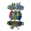 1g3iS S: Starting model for refinement |
|---|---|
| Similar structure data |
- Links
Links
- Assembly
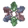
| Deposited unit | 
| ||||||||
|---|---|---|---|---|---|---|---|---|---|
| 1 |
| ||||||||
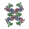
| Unit cell |
|
- Components
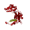
| #1: Protein | Mass: 49441.504 Da / Num. of mol.: 12 Source method: isolated from a genetically manipulated source Source: (gene. exp.)  Haemophilus influenzae (bacteria) / Strain: Rd KW20 / Gene: hslU / Plasmid: pET / Species (production host): Escherichia coli / Production host: Haemophilus influenzae (bacteria) / Strain: Rd KW20 / Gene: hslU / Plasmid: pET / Species (production host): Escherichia coli / Production host:  #2: Protein | Mass: 18903.549 Da / Num. of mol.: 12 Source method: isolated from a genetically manipulated source Source: (gene. exp.)  Haemophilus influenzae (bacteria) / Strain: Rd KW20 / Gene: hslV / Plasmid: pET / Species (production host): Escherichia coli / Production host: Haemophilus influenzae (bacteria) / Strain: Rd KW20 / Gene: hslV / Plasmid: pET / Species (production host): Escherichia coli / Production host:  #3: Chemical | ChemComp-ATP / #4: Chemical | ChemComp-LVS / Has protein modification | Y | |
|---|
-Experimental details
-Experiment
| Experiment | Method:  X-RAY DIFFRACTION / Number of used crystals: 1 X-RAY DIFFRACTION / Number of used crystals: 1 |
|---|
- Sample preparation
| Crystal | Density Matthews: 3.39 Å3/Da / Density % sol: 63.73 % | ||||||||||||||||||||||||||||||||||||||||||||||||||||||
|---|---|---|---|---|---|---|---|---|---|---|---|---|---|---|---|---|---|---|---|---|---|---|---|---|---|---|---|---|---|---|---|---|---|---|---|---|---|---|---|---|---|---|---|---|---|---|---|---|---|---|---|---|---|---|---|
| Crystal grow | Temperature: 277 K / Method: vapor diffusion, hanging drop / pH: 6 Details: PEG MONOMETHYL ETHER 2000, POTASSIUM CHLORIDE, MAGNESIUM ACETATE, CITRATE, pH 6.0, VAPOR DIFFUSION, HANGING DROP, temperature 277K | ||||||||||||||||||||||||||||||||||||||||||||||||||||||
| Crystal grow | *PLUS Temperature: 4 ℃ / pH: 7 Details: Sousa, M.C., (2000) Cell (Cambridge,Mass.), 103, 633. | ||||||||||||||||||||||||||||||||||||||||||||||||||||||
| Components of the solutions | *PLUS
|
-Data collection
| Diffraction | Mean temperature: 100 K |
|---|---|
| Diffraction source | Source:  SYNCHROTRON / Site: SYNCHROTRON / Site:  SSRL SSRL  / Beamline: BL11-1 / Wavelength: 0.965 Å / Beamline: BL11-1 / Wavelength: 0.965 Å |
| Detector | Type: ADSC QUANTUM 4 / Detector: CCD / Date: Apr 15, 2001 |
| Radiation | Monochromator: Flat mirror (vertical focusing); single crystal Si(111) bent monochromator (horizontal focusing) Protocol: SINGLE WAVELENGTH / Monochromatic (M) / Laue (L): M / Scattering type: x-ray |
| Radiation wavelength | Wavelength: 0.965 Å / Relative weight: 1 |
| Reflection | Resolution: 3.1→30 Å / Num. all: 185090 / Num. obs: 185090 / % possible obs: 92.9 % / Observed criterion σ(F): 0 / Observed criterion σ(I): 0 / Redundancy: 3.7 % / Rsym value: 0.064 / Net I/σ(I): 16.1 |
| Reflection shell | Resolution: 3.1→3.15 Å / Redundancy: 2.4 % / Mean I/σ(I) obs: 3.7 / Rsym value: 0.285 / % possible all: 87.8 |
| Reflection | *PLUS Num. measured all: 675855 / Rmerge(I) obs: 0.064 |
| Reflection shell | *PLUS % possible obs: 87.8 % / Rmerge(I) obs: 0.285 |
- Processing
| Software |
| |||||||||||||||||||||||||
|---|---|---|---|---|---|---|---|---|---|---|---|---|---|---|---|---|---|---|---|---|---|---|---|---|---|---|
| Refinement | Method to determine structure:  MOLECULAR REPLACEMENT MOLECULAR REPLACEMENTStarting model: PDB entry 1G3I Resolution: 3.1→30 Å / Cross valid method: THROUGHOUT / σ(F): 0 / Stereochemistry target values: Engh & Huber
| |||||||||||||||||||||||||
| Refinement step | Cycle: LAST / Resolution: 3.1→30 Å
| |||||||||||||||||||||||||
| Refine LS restraints |
| |||||||||||||||||||||||||
| LS refinement shell | Highest resolution: 3.1 Å
| |||||||||||||||||||||||||
| Refinement | *PLUS Rfactor obs: 0.266 / Rfactor Rfree: 0.288 / Rfactor Rwork: 0.266 | |||||||||||||||||||||||||
| Solvent computation | *PLUS | |||||||||||||||||||||||||
| Displacement parameters | *PLUS | |||||||||||||||||||||||||
| LS refinement shell | *PLUS Rfactor Rfree: 0.288 / Rfactor Rwork: 0.266 / Rfactor obs: 0.266 |
 Movie
Movie Controller






 PDBj
PDBj


