 Movie
Movie Controller

[English] 日本語
 Yorodumi
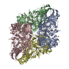
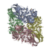
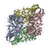
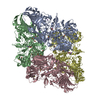
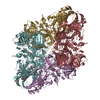
- PDB-1jz3: E. COLI (lacZ) BETA-GALACTOSIDASE-TRAPPED 2-DEOXY-GALACTOSYL ENZY... -
+ Open data

- Basic information
| Entry | Database: PDB / ID: 1jz3 | ||||||
|---|---|---|---|---|---|---|---|
| Title | E. COLI (lacZ) BETA-GALACTOSIDASE-TRAPPED 2-DEOXY-GALACTOSYL ENZYME INTERMEDIATE | ||||||
 Components Components | Beta-Galactosidase | ||||||
 Keywords Keywords | HYDROLASE / TIM BARREL (ALPHA/BETA BARREL) / JELLY-ROLL BARREL / IMMUNOGLOBULIN / BETA SUPERSANDWICH | ||||||
| Function / homology |  Function and homology information Function and homology informationalkali metal ion binding / lactose catabolic process / beta-galactosidase complex / beta-galactosidase / beta-galactosidase activity / carbohydrate binding / magnesium ion binding / identical protein binding Similarity search - Function | ||||||
| Biological species |  | ||||||
| Method |  X-RAY DIFFRACTION / SYNCHROTRON / MOLECULAR REPLACEMENT / Resolution: 1.75 Å X-RAY DIFFRACTION / SYNCHROTRON / MOLECULAR REPLACEMENT / Resolution: 1.75 Å | ||||||
 Authors Authors | Juers, D.H. / Matthews, B.W. | ||||||
 Citation Citation | Journal: Biochemistry / Year: 2001 Title: A Structural View of the Action of Escherichia Coli (Lacz) Beta-Galactosidase Authors: Juers, D.H. / Heightman, T.D. / Vasella, A. / McCarter, J.D. / Mackenzie, L. / Withers, S.G. / Matthews, B.W. #1: Journal: Protein Sci. / Year: 2000Title: High Resolution Structure of Beta-Galactosidase in a New Crystal Form Reveals Multiple Metal-Binding Sites and Provides a Structural Basis for Alpha-Complementation Authors: Juers, D.H. / Jacobson, R.H. / Wigley, D. / Zhang, X.J. / Huber, R.E. / Tronrud, D.E. / Matthews, B.W. #2: Journal: Protein Sci. / Year: 1999Title: Structural Comparisons of Tim Barrel Proteins Suggest Functional and Evolutionary Relationships between Beta-Galactosidase and Other Glycohydrolases Authors: Juers, D.H. / Huber, R.E. / Matthews, B.W. #3: Journal: Nature / Year: 1994Title: Three-Dimensional Structure of Beta-Galactosidase from E. Coli Authors: Jacobson, R.H. / Zhang, X.J. / Dubose, R.F. / Matthews, B.W. #4: Journal: J.Mol.Biol. / Year: 1992Title: Crystallization of beta-galactosidase from Escherichia coli Authors: Jacobson, R.H. / Matthews, B.W. | ||||||
| History |
|
- Structure visualization
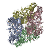
| Structure viewer | Molecule: MolmilJmol/JSmol |
|---|
- Downloads & links
-Download
| PDBx/mmCIF format | 1jz3.cif.gz | 939 KB | Display | PDBx/mmCIF format |
|---|---|---|---|---|
| PDB format | pdb1jz3.ent.gz | 745.4 KB | Display | PDB format |
| PDBx/mmJSON format | 1jz3.json.gz | Tree view | PDBx/mmJSON format | |
| Others |  Other downloads Other downloads |
-Validation report
| Arichive directory | https://data.pdbj.org/pub/pdb/validation_reports/jz/1jz3ftp://data.pdbj.org/pub/pdb/validation_reports/jz/1jz3 | HTTPS FTP |
|---|
-Related structure data
| Related structure data | 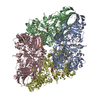 1jynC  1jyvC  1jywC  1jyxC 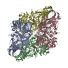 1jz2C  1jz4C  1jz5C  1jz6C  1jz7C  1jz8C  4v44C  4v45C 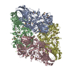 1dp0S S: Starting model for refinement C: citing same article ( |
|---|---|
| Similar structure data |


-Links
 PDBj
PDBj


- Assembly
| Deposited unit | 
| ||||||||||
|---|---|---|---|---|---|---|---|---|---|---|---|
| 1 |
| ||||||||||
| Unit cell |
|
-Components
-Protein / Sugars , 2 types, 8 molecules ABCD
| #1: Protein | Mass: 116506.266 Da / Num. of mol.: 4 Source method: isolated from a genetically manipulated source Source: (gene. exp.) #2: Sugar | ChemComp-2DG /  Type: D-saccharide, alpha linking / Mass: 164.156 Da / Num. of mol.: 4 Type: D-saccharide, alpha linking / Mass: 164.156 Da / Num. of mol.: 4Source method: isolated from a genetically manipulated source Formula: C6H12O5 |
|---|
-Non-polymers , 5 types, 4441 molecules 


| #3: Chemical | ChemComp-MG /  Mass: 24.305 Da / Num. of mol.: 17 / Source method: obtained synthetically / Formula: Mg Mass: 24.305 Da / Num. of mol.: 17 / Source method: obtained synthetically / Formula: Mg#4: Chemical | ChemComp-NA /  Mass: 22.990 Da / Num. of mol.: 16 / Source method: obtained synthetically / Formula: Na Mass: 22.990 Da / Num. of mol.: 16 / Source method: obtained synthetically / Formula: Na#5: Chemical | ChemComp-BTB /  Mass: 209.240 Da / Num. of mol.: 4 / Source method: obtained synthetically / Formula: C8H19NO5 / Comment: pH buffer*YM Mass: 209.240 Da / Num. of mol.: 4 / Source method: obtained synthetically / Formula: C8H19NO5 / Comment: pH buffer*YM#6: Chemical | ChemComp-DMS /  Mass: 78.133 Da / Num. of mol.: 110 / Source method: obtained synthetically / Formula: C2H6OS / Comment: DMSO, precipitant*YM Mass: 78.133 Da / Num. of mol.: 110 / Source method: obtained synthetically / Formula: C2H6OS / Comment: DMSO, precipitant*YM#7: Water | ChemComp-HOH / | Mass: 18.015 Da / Num. of mol.: 4294 / Source method: isolated from a natural source / Formula: H2O |
|---|
-Details
| Has protein modification | Y |
|---|
-Experimental details
-Experiment
| Experiment | Method: X-RAY DIFFRACTION / Number of used crystals: 1 |
|---|
- Sample preparation
| Crystal | Density Matthews: 2.7 Å3/Da / Density % sol: 55 % | |||||||||||||||||||||||||||||||||||||||||||||||||||||||||||||||||||||||||||||
|---|---|---|---|---|---|---|---|---|---|---|---|---|---|---|---|---|---|---|---|---|---|---|---|---|---|---|---|---|---|---|---|---|---|---|---|---|---|---|---|---|---|---|---|---|---|---|---|---|---|---|---|---|---|---|---|---|---|---|---|---|---|---|---|---|---|---|---|---|---|---|---|---|---|---|---|---|---|---|
| Crystal grow | Temperature: 288 K / Method: vapor diffusion, hanging drop / pH: 6.5 Details: Bis-Tris, PEG 8000, MgCl2, NaCl, DTT, pH 6.5, VAPOR DIFFUSION, HANGING DROP at 288K, pH 6.50 | |||||||||||||||||||||||||||||||||||||||||||||||||||||||||||||||||||||||||||||
| Crystal grow | *PLUS pH: 6.5 / Method: vapor diffusionDetails: used macroseeding, Juers, D.H., (2000) Protein Sci., 9, 1685. | |||||||||||||||||||||||||||||||||||||||||||||||||||||||||||||||||||||||||||||
| Components of the solutions | *PLUS
|
-Data collection
| Diffraction | Mean temperature: 100 K | |||||||||
|---|---|---|---|---|---|---|---|---|---|---|
| Diffraction source | Source: SYNCHROTRON / Site: ALS  / Beamline: 5.0.1 / Wavelength: 1 / Wavelength: 1 Å / Beamline: 5.0.1 / Wavelength: 1 / Wavelength: 1 Å | |||||||||
| Detector | Type: ADSC QUANTUM 4 / Detector: CCD / Date: Mar 13, 1998 | |||||||||
| Radiation | Protocol: SINGLE WAVELENGTH / Monochromatic (M) / Laue (L): M / Scattering type: x-ray | |||||||||
| Radiation wavelength |
| |||||||||
| Reflection | Resolution: 1.75→16 Å / Num. all: 470782 / Num. obs: 470782 / % possible obs: 93.8 % / Redundancy: 3.3 % / Biso Wilson estimate: 15.6 Å2 / Rmerge(I) obs: 0.061 / Net I/σ(I): 14 | |||||||||
| Reflection shell | Highest resolution: 1.75 Å / Rmerge(I) obs: 0.46 / Mean I/σ(I) obs: 2.2 / % possible all: 87.5 | |||||||||
| Reflection | *PLUS % possible obs: 94 % / Redundancy: 3.3 % / Rmerge(I) obs: 0.061 | |||||||||
| Reflection shell | *PLUS Rmerge(I) obs: 0.46 / Mean I/σ(I) obs: 2.2 |
- Processing
| Software |
| ||||||||||||||||||||||||||||||||||||||||||||||||||
|---|---|---|---|---|---|---|---|---|---|---|---|---|---|---|---|---|---|---|---|---|---|---|---|---|---|---|---|---|---|---|---|---|---|---|---|---|---|---|---|---|---|---|---|---|---|---|---|---|---|---|---|
| Refinement | Method to determine structure: MOLECULAR REPLACEMENT Starting model: 1DP0 Resolution: 1.75→16 Å / Isotropic thermal model: ISOTROPIC / Cross valid method: THROUGHOUT / Stereochemistry target values: TNT
| ||||||||||||||||||||||||||||||||||||||||||||||||||
| Solvent computation | Solvent model: BABINET PRINCIPLE / Bsol: 115 Å2 / ksol: 0.7 e/Å3 | ||||||||||||||||||||||||||||||||||||||||||||||||||
| Displacement parameters | Biso mean: 23.6 Å2
| ||||||||||||||||||||||||||||||||||||||||||||||||||
| Refinement step | Cycle: LAST / Resolution: 1.75→16 Å
| ||||||||||||||||||||||||||||||||||||||||||||||||||
| Refine LS restraints |
| ||||||||||||||||||||||||||||||||||||||||||||||||||
| Refinement | *PLUS Rfactor obs: 0.158 / Rfactor Rfree: 0.222 / Rfactor Rwork: 0.158 | ||||||||||||||||||||||||||||||||||||||||||||||||||
| Solvent computation | *PLUS | ||||||||||||||||||||||||||||||||||||||||||||||||||
| Displacement parameters | *PLUS | ||||||||||||||||||||||||||||||||||||||||||||||||||
| Refine LS restraints | *PLUS
|
