 Movie
Movie Controller


+ Open data

- Basic information
| Entry | Database: PDB / ID: 6cvm | ||||||
|---|---|---|---|---|---|---|---|
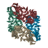
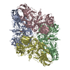
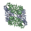
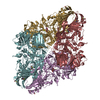
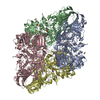
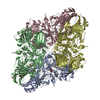
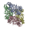
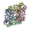
| Title | Atomic resolution cryo-EM structure of beta-galactosidase | ||||||
 Components Components | Beta-galactosidase | ||||||
 Keywords Keywords | HYDROLASE / drift correction / radiation damage / drug discovery / precision medicine / computer-aided drug discovery | ||||||
| Function / homology |  Function and homology information Function and homology informationalkali metal ion binding / lactose catabolic process / beta-galactosidase complex / beta-galactosidase / beta-galactosidase activity / carbohydrate binding / magnesium ion binding / identical protein binding Similarity search - Function | ||||||
| Biological species |  | ||||||
| Method | ELECTRON MICROSCOPY / single particle reconstruction / cryo EM / Resolution: 1.9 Å | ||||||
 Authors Authors | Subramaniam, S. / Bartesaghi, A. / Banerjee, S. / Zhu, X. / Milne, J.L.S. | ||||||
 Citation Citation | Journal: Structure / Year: 2018 Title: Atomic Resolution Cryo-EM Structure of β-Galactosidase. Authors: Alberto Bartesaghi / Cecilia Aguerrebere / Veronica Falconieri / Soojay Banerjee / Lesley A Earl / Xing Zhu / Nikolaus Grigorieff / Jacqueline L S Milne / Guillermo Sapiro / Xiongwu Wu / Sriram Subramaniam /  Abstract: The advent of direct electron detectors has enabled the routine use of single-particle cryo-electron microscopy (EM) approaches to determine structures of a variety of protein complexes at near- ...The advent of direct electron detectors has enabled the routine use of single-particle cryo-electron microscopy (EM) approaches to determine structures of a variety of protein complexes at near-atomic resolution. Here, we report the development of methods to account for local variations in defocus and beam-induced drift, and the implementation of a data-driven dose compensation scheme that significantly improves the extraction of high-resolution information recorded during exposure of the specimen to the electron beam. These advances enable determination of a cryo-EM density map for β-galactosidase bound to the inhibitor phenylethyl β-D-thiogalactopyranoside where the ordered regions are resolved at a level of detail seen in X-ray maps at ∼ 1.5 Å resolution. Using this density map in conjunction with constrained molecular dynamics simulations provides a measure of the local flexibility of the non-covalently bound inhibitor and offers further opportunities for structure-guided inhibitor design. | ||||||
| History |
|
- Structure visualization
| Movie |
Movie viewer |
|---|---|
| Structure viewer | Molecule: MolmilJmol/JSmol |
- Downloads & links
-Download
| PDBx/mmCIF format | 6cvm.cif.gz | 837.8 KB | Display | PDBx/mmCIF format |
|---|---|---|---|---|
| PDB format | pdb6cvm.ent.gz | 669.4 KB | Display | PDB format |
| PDBx/mmJSON format | 6cvm.json.gz | Tree view | PDBx/mmJSON format | |
| Others |  Other downloads Other downloads |
-Validation report
| Arichive directory | https://data.pdbj.org/pub/pdb/validation_reports/cv/6cvmftp://data.pdbj.org/pub/pdb/validation_reports/cv/6cvm | HTTPS FTP |
|---|
-Related structure data
| Related structure data | 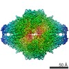 7770MC M: map data used to model this data C: citing same article ( |
|---|---|
| Similar structure data | |
| Experimental dataset #1 | Data reference: 10.6019/EMPIAR-10061 / Data set type: EMPIAR / Metadata reference: 10.6019/EMPIAR-10061 |


-Links
 PDBj
PDBj


- Assembly
| Deposited unit | 
|
|---|---|
| 1 |
|
-Components
| #1: Protein | Mass: 116181.031 Da / Num. of mol.: 4 / Source method: isolated from a natural source / Source: (natural) #2: Sugar | ChemComp-PTQ /   Type: D-saccharide / Mass: 300.371 Da / Num. of mol.: 4 Type: D-saccharide / Mass: 300.371 Da / Num. of mol.: 4Source method: isolated from a genetically manipulated source Formula: C14H20O5S #3: Chemical | ChemComp-MG /   Mass: 24.305 Da / Num. of mol.: 8 / Source method: obtained synthetically / Formula: Mg Mass: 24.305 Da / Num. of mol.: 8 / Source method: obtained synthetically / Formula: Mg#4: Chemical | ChemComp-NA /   Mass: 22.990 Da / Num. of mol.: 8 / Source method: obtained synthetically / Formula: Na Mass: 22.990 Da / Num. of mol.: 8 / Source method: obtained synthetically / Formula: Na#5: Water | ChemComp-HOH / |  Mass: 18.015 Da / Num. of mol.: 4194 / Source method: isolated from a natural source / Formula: H2O Mass: 18.015 Da / Num. of mol.: 4194 / Source method: isolated from a natural source / Formula: H2O |
|---|
-Experimental details
-Experiment
| Experiment | Method: ELECTRON MICROSCOPY |
|---|---|
| EM experiment | Aggregation state: PARTICLE / 3D reconstruction method: single particle reconstruction |
- Sample preparation
| Component | Name: Escherichia coli beta-galactosidase bound to phenylethyl beta-D-thiogalactopyranoside (PETG) Type: COMPLEX / Entity ID: #1 / Source: NATURAL |
|---|---|
| Molecular weight | Value: 0.465 MDa / Experimental value: NO |
| Source (natural) | Organism: |
| Buffer solution | pH: 8 Details: 25 mM Tris, pH 8.0, 50 mM NaCl, 2 mM MgCl2, 1.0 mM TCEP |
| Specimen | Conc.: 2.3 mg/ml / Embedding applied: NO / Shadowing applied: NO / Staining applied: NO / Vitrification applied: YES |
| Specimen support | Details: plasma cleaned / Grid material: COPPER / Grid mesh size: 200 divisions/in. / Grid type: Quantifoil R2/2 |
| Vitrification | Instrument: LEICA EM GP / Cryogen name: ETHANE / Humidity: 90 % / Chamber temperature: 90.15 K / Details: Blot for 2 seconds before plunging. |
- Electron microscopy imaging
| Experimental equipment |  Model: Titan Krios / Image courtesy: FEI Company |
|---|---|
| Microscopy | Model: FEI TITAN KRIOS / Details: Parallel beam illumination |
| Electron gun | Electron source:  FIELD EMISSION GUN / Accelerating voltage: 300 kV / Illumination mode: FLOOD BEAM FIELD EMISSION GUN / Accelerating voltage: 300 kV / Illumination mode: FLOOD BEAM |
| Electron lens | Mode: BRIGHT FIELD / Nominal magnification: 215000 X / Calibrated magnification: 215000 X / Nominal defocus min: 600 nm / Calibrated defocus max: 2000 nm / Cs: 2.7 mm |
| Specimen holder | Cryogen: NITROGEN / Specimen holder model: FEI TITAN KRIOS AUTOGRID HOLDER / Temperature (max): 79.8 K / Temperature (min): 79.6 K |
| Image recording | Average exposure time: 7.6 sec. / Electron dose: 45 e/Å2 / Detector mode: SUPER-RESOLUTION / Film or detector model: GATAN K2 QUANTUM (4k x 4k) / Num. of real images: 1539 / Details: Raw micrographs are available from EMPIAR-10061. |
| EM imaging optics | Energyfilter name: GIF Quantum LS / Energyfilter upper: 20 eV / Energyfilter lower: 0 eV |
| Image scans | Movie frames/image: 38 / Used frames/image: 1-38 |
- Processing
| Software | Name: PHENIX / Version: 1.13_2998: / Classification: refinement | ||||||||||||||||||||||||||||||
|---|---|---|---|---|---|---|---|---|---|---|---|---|---|---|---|---|---|---|---|---|---|---|---|---|---|---|---|---|---|---|---|
| EM software |
| ||||||||||||||||||||||||||||||
| CTF correction | Type: PHASE FLIPPING AND AMPLITUDE CORRECTION | ||||||||||||||||||||||||||||||
| Particle selection | Num. of particles selected: 298715 | ||||||||||||||||||||||||||||||
| Symmetry | Point symmetry: D2 (2x2 fold dihedral) | ||||||||||||||||||||||||||||||
| 3D reconstruction | Resolution: 1.9 Å / Resolution method: FSC 0.143 CUT-OFF / Num. of particles: 150321 / Algorithm: FOURIER SPACE / Symmetry type: POINT | ||||||||||||||||||||||||||||||
| Atomic model building | Protocol: FLEXIBLE FIT / Space: REAL | ||||||||||||||||||||||||||||||
| Refine LS restraints |
|
