 Movie
Movie Controller

[English] 日本語
 Yorodumi
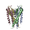
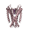
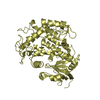
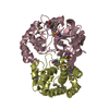
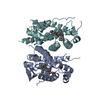
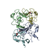
- PDB-1jvm: KCSA POTASSIUM CHANNEL WITH TBA (TETRABUTYLAMMONIUM) AND RUBIDIUM -
+ Open data

- Basic information
| Entry | Database: PDB / ID: 1jvm | ||||||
|---|---|---|---|---|---|---|---|
| Title | KCSA POTASSIUM CHANNEL WITH TBA (TETRABUTYLAMMONIUM) AND RUBIDIUM | ||||||
 Components Components | Voltage-gated potassium channel | ||||||
 Keywords Keywords | MEMBRANE PROTEIN / POTASSIUM CHANNEL / METAL TRANSPORT | ||||||
| Function / homology |  Function and homology information Function and homology informationaction potential / voltage-gated potassium channel activity / voltage-gated potassium channel complex / identical protein binding Similarity search - Function | ||||||
| Biological species |  Streptomyces lividans (bacteria) Streptomyces lividans (bacteria) | ||||||
| Method |  X-RAY DIFFRACTION / SYNCHROTRON / MOLECULAR REPLACEMENT / Resolution: 2.8 Å X-RAY DIFFRACTION / SYNCHROTRON / MOLECULAR REPLACEMENT / Resolution: 2.8 Å | ||||||
 Authors Authors | Morais-Cabral, J.H. / Zhou, Y. / MacKinnon, R. | ||||||
 Citation Citation | Journal: Nature / Year: 2001 Title: Energetic optimization of ion conduction rate by the K+ selectivity filter. Authors: Morais-Cabral, J.H. / Zhou, Y. / MacKinnon, R. | ||||||
| History |
|
- Structure visualization
| Structure viewer | Molecule: MolmilJmol/JSmol |
|---|
- Downloads & links
-Download
| PDBx/mmCIF format | 1jvm.cif.gz | 84.2 KB | Display | PDBx/mmCIF format |
|---|---|---|---|---|
| PDB format | pdb1jvm.ent.gz | 62.8 KB | Display | PDB format |
| PDBx/mmJSON format | 1jvm.json.gz | Tree view | PDBx/mmJSON format | |
| Others |  Other downloads Other downloads |
-Validation report
| Arichive directory | https://data.pdbj.org/pub/pdb/validation_reports/jv/1jvmftp://data.pdbj.org/pub/pdb/validation_reports/jv/1jvm | HTTPS FTP |
|---|
-Related structure data
| Related structure data |  1bl8S S: Starting model for refinement |
|---|---|
| Similar structure data |


-Links
 PDBj
PDBj


- Assembly
| Deposited unit | 
| ||||||||
|---|---|---|---|---|---|---|---|---|---|
| 1 |
| ||||||||
| 2 | 
| ||||||||
| Unit cell |
|
-Components
| #1: Protein | Mass: 13358.756 Da / Num. of mol.: 4 / Fragment: Residues 1-125 / Mutation: P2A,L90C Source method: isolated from a genetically manipulated source Source: (gene. exp.) Streptomyces lividans (bacteria) / Plasmid: pQE-60 / Production host: #2: Chemical |   Mass: 85.468 Da / Num. of mol.: 3 / Source method: obtained synthetically / Formula: Rb Mass: 85.468 Da / Num. of mol.: 3 / Source method: obtained synthetically / Formula: Rb#3: Chemical | ChemComp-TBA / |   Mass: 242.464 Da / Num. of mol.: 1 / Source method: obtained synthetically / Formula: C16H36N Mass: 242.464 Da / Num. of mol.: 1 / Source method: obtained synthetically / Formula: C16H36N#4: Water | ChemComp-HOH / |  Mass: 18.015 Da / Num. of mol.: 1 / Source method: isolated from a natural source / Formula: H2O Mass: 18.015 Da / Num. of mol.: 1 / Source method: isolated from a natural source / Formula: H2O |
|---|
-Experimental details
-Experiment
| Experiment | Method: X-RAY DIFFRACTION / Number of used crystals: 1 |
|---|
- Sample preparation
| Crystal | Density Matthews: 3.86 Å3/Da / Density % sol: 68.14 % | ||||||||||||||||||||||||||||||||||||||||||||||||||||||||
|---|---|---|---|---|---|---|---|---|---|---|---|---|---|---|---|---|---|---|---|---|---|---|---|---|---|---|---|---|---|---|---|---|---|---|---|---|---|---|---|---|---|---|---|---|---|---|---|---|---|---|---|---|---|---|---|---|---|
| Crystal grow | Temperature: 293 K / Method: vapor diffusion, sitting drop / pH: 7.5 Details: PEG400,calcium chloride, Hepes, potassium chloride, pH 7.5, VAPOR DIFFUSION, SITTING DROP, temperature 293K | ||||||||||||||||||||||||||||||||||||||||||||||||||||||||
| Crystal grow | *PLUS Temperature: 20 ℃ | ||||||||||||||||||||||||||||||||||||||||||||||||||||||||
| Components of the solutions | *PLUS
|
-Data collection
| Diffraction | Mean temperature: 100 K |
|---|---|
| Diffraction source | Source: SYNCHROTRON / Site: ESRF  / Beamline: ID13 / Wavelength: 0.964 Å / Beamline: ID13 / Wavelength: 0.964 Å |
| Detector | Type: MARRESEARCH / Detector: IMAGE PLATE / Date: Jun 25, 2000 |
| Radiation | Protocol: SINGLE WAVELENGTH / Monochromatic (M) / Laue (L): M / Scattering type: x-ray |
| Radiation wavelength | Wavelength: 0.964 Å / Relative weight: 1 |
| Reflection | Resolution: 2.7→50 Å / Num. all: 22300 / Num. obs: 14799 / % possible obs: 0.664 % / Observed criterion σ(F): 1 / Observed criterion σ(I): 1 / Redundancy: 2.5 % / Rmerge(I) obs: 0.048 / Net I/σ(I): 21 |
| Reflection shell | Resolution: 2.7→2.8 Å / Rmerge(I) obs: 0.088 / % possible all: 0.167 |
| Reflection | *PLUS Lowest resolution: 50 Å / Rmerge(I) obs: 0.048 |
| Reflection shell | *PLUS Rmerge(I) obs: 0.088 |
- Processing
| Software |
| |||||||||||||||||||||||||
|---|---|---|---|---|---|---|---|---|---|---|---|---|---|---|---|---|---|---|---|---|---|---|---|---|---|---|
| Refinement | Method to determine structure: MOLECULAR REPLACEMENT Starting model: PDB Entry 1BL8 Resolution: 2.8→30 Å / Isotropic thermal model: isotropic / Cross valid method: THROUGHOUT / σ(F): 0 / Stereochemistry target values: Engh & Huber
| |||||||||||||||||||||||||
| Refinement step | Cycle: LAST / Resolution: 2.8→30 Å
| |||||||||||||||||||||||||
| Refine LS restraints |
| |||||||||||||||||||||||||
| Refinement | *PLUS % reflection Rfree: 10 % / Rfactor all: 0.29 / Rfactor obs: 0.281 / Rfactor Rfree: 0.3021 / Rfactor Rwork: 0.281 | |||||||||||||||||||||||||
| Solvent computation | *PLUS | |||||||||||||||||||||||||
| Displacement parameters | *PLUS |
