 Movie
Movie Controller


+ Open data

- Basic information
| Entry | Database: PDB / ID: 1ju3 | ||||||
|---|---|---|---|---|---|---|---|
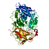
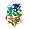
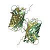
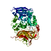
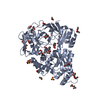
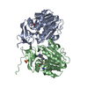
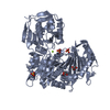
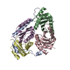
| Title | BACTERIAL COCAINE ESTERASE COMPLEX WITH TRANSITION STATE ANALOG | ||||||
 Components Components | cocaine esterase | ||||||
 Keywords Keywords | HYDROLASE / alpha/beta hydrolase | ||||||
| Function / homology |  Function and homology information Function and homology informationcocaine esterase / cocaine catabolic process / dipeptidyl-peptidase activity / carboxylic ester hydrolase activity / cytoplasm Similarity search - Function | ||||||
| Biological species |  Rhodococcus sp. MB1 (bacteria) Rhodococcus sp. MB1 (bacteria) | ||||||
| Method |  X-RAY DIFFRACTION / SYNCHROTRON / MOLECULAR REPLACEMENT / Resolution: 1.58 Å X-RAY DIFFRACTION / SYNCHROTRON / MOLECULAR REPLACEMENT / Resolution: 1.58 Å | ||||||
 Authors Authors | Larsen, N.A. / Turner, J.M. / Stevens, J. / Rosser, S.J. / Basran, A. / Lerner, R.A. / Bruce, N.C. / Wilson, I.A. | ||||||
 Citation Citation | Journal: Nat.Struct.Biol. / Year: 2002 Title: Crystal structure of a bacterial cocaine esterase. Authors: Larsen, N.A. / Turner, J.M. / Stevens, J. / Rosser, S.J. / Basran, A. / Lerner, R.A. / Bruce, N.C. / Wilson, I.A. | ||||||
| History |
|
- Structure visualization
| Structure viewer | Molecule: MolmilJmol/JSmol |
|---|
- Downloads & links
-Download
| PDBx/mmCIF format | 1ju3.cif.gz | 133.8 KB | Display | PDBx/mmCIF format |
|---|---|---|---|---|
| PDB format | pdb1ju3.ent.gz | 102.3 KB | Display | PDB format |
| PDBx/mmJSON format | 1ju3.json.gz | Tree view | PDBx/mmJSON format | |
| Others |  Other downloads Other downloads |
-Validation report
| Arichive directory | https://data.pdbj.org/pub/pdb/validation_reports/ju/1ju3ftp://data.pdbj.org/pub/pdb/validation_reports/ju/1ju3 | HTTPS FTP |
|---|
-Related structure data
| Related structure data | 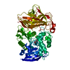 1ju4SC S: Starting model for refinement C: citing same article ( |
|---|---|
| Similar structure data |

-Links
 PDBj
PDBj- Assembly
| Deposited unit | 
| ||||||||
|---|---|---|---|---|---|---|---|---|---|
| 1 |
| ||||||||
| Unit cell |
|
-Components
| #1: Protein | Mass: 63351.637 Da / Num. of mol.: 1 Source method: isolated from a genetically manipulated source Source: (gene. exp.) Rhodococcus sp. MB1 (bacteria) / Production host: |
|---|---|
| #2: Chemical | ChemComp-PBC /   Mass: 121.930 Da / Num. of mol.: 1 / Source method: obtained synthetically / Formula: C6H7BO2 Mass: 121.930 Da / Num. of mol.: 1 / Source method: obtained synthetically / Formula: C6H7BO2 |
| #3: Water | ChemComp-HOH /  Mass: 18.015 Da / Num. of mol.: 544 / Source method: isolated from a natural source / Formula: H2O Mass: 18.015 Da / Num. of mol.: 544 / Source method: isolated from a natural source / Formula: H2O |
| Has protein modification | Y |
-Experimental details
-Experiment
| Experiment | Method: X-RAY DIFFRACTION / Number of used crystals: 2 |
|---|
- Sample preparation
| Crystal | Density Matthews: 2.84 Å3/Da / Density % sol: 56.66 % | ||||||||||||||||||||||||||||||||||||||||||
|---|---|---|---|---|---|---|---|---|---|---|---|---|---|---|---|---|---|---|---|---|---|---|---|---|---|---|---|---|---|---|---|---|---|---|---|---|---|---|---|---|---|---|---|
| Crystal grow | Temperature: 295.5 K / Method: vapor diffusion, sitting drop / pH: 7.5 Details: 10mM DTT, 10mM Tris pH 7.5, 25 mM NaCl, 1.6 M Ammonium Sulfate, VAPOR DIFFUSION, SITTING DROP, temperature 22.5K | ||||||||||||||||||||||||||||||||||||||||||
| Crystal | *PLUS Density % sol: 53 % | ||||||||||||||||||||||||||||||||||||||||||
| Crystal grow | *PLUS Method: microdialysis | ||||||||||||||||||||||||||||||||||||||||||
| Components of the solutions | *PLUS
|
-Data collection
| Diffraction |
| |||||||||||||||
|---|---|---|---|---|---|---|---|---|---|---|---|---|---|---|---|---|
| Diffraction source |
| |||||||||||||||
| Detector |
| |||||||||||||||
| Radiation |
| |||||||||||||||
| Radiation wavelength | Wavelength: 0.965 Å / Relative weight: 1 | |||||||||||||||
| Reflection | Resolution: 1.58→20 Å / Num. all: 90349 / Num. obs: 90349 / % possible obs: 90 % / Observed criterion σ(F): 2 / Observed criterion σ(I): 2 / Rmerge(I) obs: 0.064 | |||||||||||||||
| Reflection shell | Resolution: 1.58→1.61 Å / Rmerge(I) obs: 0.514 / % possible all: 71.2 | |||||||||||||||
| Reflection | *PLUS Lowest resolution: 20 Å / Redundancy: 5.4 % | |||||||||||||||
| Reflection shell | *PLUS % possible obs: 71.2 % / Mean I/σ(I) obs: 2.2 |

- Processing
| Software |
| |||||||||||||||||||||
|---|---|---|---|---|---|---|---|---|---|---|---|---|---|---|---|---|---|---|---|---|---|---|
| Refinement | Method to determine structure: MOLECULAR REPLACEMENT Starting model: PDB ENTRY 1JU4, cocE enzyme Resolution: 1.58→20 Å / σ(F): 0 / Stereochemistry target values: Engh & Huber
| |||||||||||||||||||||
| Refine analyze | Luzzati coordinate error obs: 0.1881 Å / Luzzati d res low obs: 20 Å / Luzzati sigma a obs: 0.1312 Å | |||||||||||||||||||||
| Refinement step | Cycle: LAST / Resolution: 1.58→20 Å
| |||||||||||||||||||||
| Refine LS restraints |
| |||||||||||||||||||||
| Software | *PLUS Name: CNS / Classification: refinement | |||||||||||||||||||||
| Refinement | *PLUS Lowest resolution: 20 Å / σ(F): 0 / % reflection Rfree: 5 % | |||||||||||||||||||||
| Solvent computation | *PLUS | |||||||||||||||||||||
| Displacement parameters | *PLUS | |||||||||||||||||||||
| Refine LS restraints | *PLUS
|
