 Movie
Movie Controller

[English] 日本語
 Yorodumi
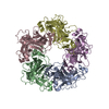
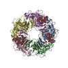
- PDB-1hnu: CRYSTAL STRUCTURE OF PEROXISOMAL DELTA3-DELTA2-ENOYL-COA ISOMERAS... -
+ Open data

- Basic information
| Entry | Database: PDB / ID: 1hnu | ||||||
|---|---|---|---|---|---|---|---|
| Title | CRYSTAL STRUCTURE OF PEROXISOMAL DELTA3-DELTA2-ENOYL-COA ISOMERASE FROM SACCHAROMYCES CEREVISIAE | ||||||
 Components Components | D3,D2-ENOYL COA ISOMERASE ECI1 | ||||||
 Keywords Keywords | ISOMERASE / alpha/beta | ||||||
| Function / homology |  Function and homology information Function and homology informationBeta-oxidation of very long chain fatty acids / Delta3-Delta2-enoyl-CoA isomerase / delta(3)-delta(2)-enoyl-CoA isomerase activity / Peroxisomal protein import / fatty acid beta-oxidation / peroxisomal matrix / peroxisome Similarity search - Function | ||||||
| Biological species |  | ||||||
| Method |  X-RAY DIFFRACTION / SYNCHROTRON / MAD / Resolution: 2.15 Å X-RAY DIFFRACTION / SYNCHROTRON / MAD / Resolution: 2.15 Å | ||||||
 Authors Authors | Mursula, A.M. / van Aalten, D.M.F. / Hiltunen, J.K. / Wierenga, R.K. | ||||||
 Citation Citation | Journal: J.Mol.Biol. / Year: 2001 Title: The crystal structure of delta(3)-delta(2)-enoyl-CoA isomerase. Authors: Mursula, A.M. / van Aalten, D.M. / Hiltunen, J.K. / Wierenga, R.K. #1: Journal: Acta Crystallogr.,Sect.D / Year: 2000Title: Crystallization and X-ray Diffraction Analysis of Peroxisomal delta3-delta2-enoyl-CoA Isomerase from Saccharomyces cerevisiae Authors: Mursula, A.M. / van Aalten, D.M. / Modis, Y. / Hiltunen, J.K. / Wierenga, R.K. | ||||||
| History |
|
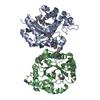
- Structure visualization
| Structure viewer | Molecule: MolmilJmol/JSmol |
|---|
- Downloads & links
-Download
| PDBx/mmCIF format | 1hnu.cif.gz | 68.1 KB | Display | PDBx/mmCIF format |
|---|---|---|---|---|
| PDB format | pdb1hnu.ent.gz | 50.6 KB | Display | PDB format |
| PDBx/mmJSON format | 1hnu.json.gz | Tree view | PDBx/mmJSON format | |
| Others |  Other downloads Other downloads |
-Validation report
| Summary document | 1hnu_validation.pdf.gz | 438.6 KB | Display | wwPDB validaton report |
|---|---|---|---|---|
| Full document | 1hnu_full_validation.pdf.gz | 444.8 KB | Display | |
| Data in XML | 1hnu_validation.xml.gz | 14.4 KB | Display | |
| Data in CIF | 1hnu_validation.cif.gz | 20.4 KB | Display | |
| Arichive directory | https://data.pdbj.org/pub/pdb/validation_reports/hn/1hnuftp://data.pdbj.org/pub/pdb/validation_reports/hn/1hnu | HTTPS FTP |
-Related structure data


-Links
 PDBj
PDBj
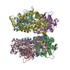
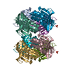
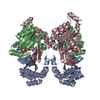
- Assembly
| Deposited unit | 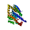
| |||||||||||||||
|---|---|---|---|---|---|---|---|---|---|---|---|---|---|---|---|---|
| 1 | x 6
| |||||||||||||||
| 2 | 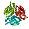
| |||||||||||||||
| Unit cell |
| |||||||||||||||
| Components on special symmetry positions |
|
-Components
| #1: Protein | Mass: 31718.369 Da / Num. of mol.: 1 Source method: isolated from a genetically manipulated source Source: (gene. exp.) Plasmid: PET3A / Production host:  References: UniProt: Q05871, Delta3-Delta2-enoyl-CoA isomerase | ||||
|---|---|---|---|---|---|
| #2: Chemical |   Mass: 250.205 Da / Num. of mol.: 2 / Source method: obtained synthetically / Formula: O4Re Mass: 250.205 Da / Num. of mol.: 2 / Source method: obtained synthetically / Formula: O4Re#3: Chemical |   Mass: 62.068 Da / Num. of mol.: 2 / Source method: obtained synthetically / Formula: C2H6O2 Mass: 62.068 Da / Num. of mol.: 2 / Source method: obtained synthetically / Formula: C2H6O2#4: Water | ChemComp-HOH / |  Mass: 18.015 Da / Num. of mol.: 194 / Source method: isolated from a natural source / Formula: H2O Mass: 18.015 Da / Num. of mol.: 194 / Source method: isolated from a natural source / Formula: H2O |
-Experimental details
-Experiment
| Experiment | Method: X-RAY DIFFRACTION / Number of used crystals: 1 |
|---|
- Sample preparation
| Crystal | Density Matthews: 3.78 Å3/Da / Density % sol: 67.48 % | ||||||||||||||||||||
|---|---|---|---|---|---|---|---|---|---|---|---|---|---|---|---|---|---|---|---|---|---|
| Crystal grow | Temperature: 295 K / Method: vapor diffusion, hanging drop / pH: 5.6 Details: MES, ammonium sulfate, 1,4-dioxane, pH 5.6, VAPOR DIFFUSION, HANGING DROP, temperature 295K | ||||||||||||||||||||
| Crystal grow | *PLUS Method: unknown | ||||||||||||||||||||
| Components of the solutions | *PLUS
|
-Data collection
| Diffraction | Mean temperature: 100 K | |||||||||||||||
|---|---|---|---|---|---|---|---|---|---|---|---|---|---|---|---|---|
| Diffraction source | Source: SYNCHROTRON / Site: EMBL/DESY, HAMBURG  / Beamline: BW7A / Wavelength: 1.1765, 1.1773, 1.1697, 1.1836 / Beamline: BW7A / Wavelength: 1.1765, 1.1773, 1.1697, 1.1836 | |||||||||||||||
| Detector | Type: MARRESEARCH / Detector: CCD / Date: Nov 26, 1999 | |||||||||||||||
| Radiation | Monochromator: synchrotron / Protocol: MAD / Monochromatic (M) / Laue (L): M / Scattering type: x-ray | |||||||||||||||
| Radiation wavelength |
| |||||||||||||||
| Reflection | Resolution: 2.15→30 Å / Num. all: 27191 / Num. obs: 27191 / % possible obs: 99.5 % / Observed criterion σ(F): 0 / Observed criterion σ(I): -3 / Redundancy: 4.8 % / Biso Wilson estimate: 40.97 Å2 / Rmerge(I) obs: 0.053 / Net I/σ(I): 16 | |||||||||||||||
| Reflection shell | Resolution: 2.15→2.23 Å / Redundancy: 4.8 % / Rmerge(I) obs: 0.474 / Mean I/σ(I) obs: 2.9 / Num. unique all: 2651 / % possible all: 99.7 | |||||||||||||||
| Reflection | *PLUS Lowest resolution: 30 Å | |||||||||||||||
| Reflection shell | *PLUS % possible obs: 99.7 % / Num. unique obs: 2651 |
- Processing
| Software |
| ||||||||||||||||||||
|---|---|---|---|---|---|---|---|---|---|---|---|---|---|---|---|---|---|---|---|---|---|
| Refinement | Method to determine structure: MAD / Resolution: 2.15→20 Å / Isotropic thermal model: individual isotropic / σ(F): 0 / σ(I): 0 / Stereochemistry target values: Engh & Huber
| ||||||||||||||||||||
| Displacement parameters | Biso mean: 44.95 Å2 | ||||||||||||||||||||
| Refinement step | Cycle: LAST / Resolution: 2.15→20 Å
| ||||||||||||||||||||
| Refine LS restraints |
| ||||||||||||||||||||
| LS refinement shell | Resolution: 2.15→2.25 Å
| ||||||||||||||||||||
| Software | *PLUS Name: REFMAC / Classification: refinement | ||||||||||||||||||||
| Refinement | *PLUS Lowest resolution: 20 Å / σ(F): 0 | ||||||||||||||||||||
| Solvent computation | *PLUS | ||||||||||||||||||||
| Displacement parameters | *PLUS | ||||||||||||||||||||
| Refine LS restraints | *PLUS
|
