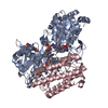
 Movie
Movie Controller


+ Open data

- Basic information
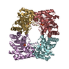
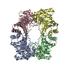
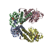
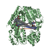
| Entry | Database: PDB / ID: 1hg3 | ||||||
|---|---|---|---|---|---|---|---|
| Title | Crystal structure of tetrameric TIM from Pyrococcus woesei. | ||||||
 Components Components | TRIOSEPHOSPHATE ISOMERASE | ||||||
 Keywords Keywords | ISOMERASE / THERMOSTABILITY / PYROCOCCUS / TRIOSEPHOSPHATE ISOMERASE / TETRAMERIC | ||||||
| Function / homology |  Function and homology information Function and homology informationtriose-phosphate isomerase / triose-phosphate isomerase activity / glyceraldehyde-3-phosphate biosynthetic process / glycerol catabolic process / gluconeogenesis / glycolytic process / cytosol / cytoplasm Similarity search - Function | ||||||
| Biological species |   PYROCOCCUS WOESEI (archaea) PYROCOCCUS WOESEI (archaea) | ||||||
| Method |  X-RAY DIFFRACTION / SYNCHROTRON / MAD / Resolution: 2.7 Å X-RAY DIFFRACTION / SYNCHROTRON / MAD / Resolution: 2.7 Å | ||||||
 Authors Authors | Walden, H. / Bell, G.S. / Russell, R.J.M. / Siebers, B. / Hensel, R. / Taylor, G.L. | ||||||
 Citation Citation | Journal: J.Mol.Biol. / Year: 2001 Title: Tiny Tim: A Small, Tetrameric, Hyperthermostable Triosephosphate Isomerase Authors: Walden, H. / Bell, G.S. / Russell, R.J.M. / Siebers, B. / Hensel, R. / Taylor, G.L. | ||||||
| History |
|
- Structure visualization
| Structure viewer | Molecule: MolmilJmol/JSmol |
|---|
- Downloads & links
-Download
| PDBx/mmCIF format | 1hg3.cif.gz | 307.1 KB | Display | PDBx/mmCIF format |
|---|---|---|---|---|
| PDB format | pdb1hg3.ent.gz | 258.2 KB | Display | PDB format |
| PDBx/mmJSON format | 1hg3.json.gz | Tree view | PDBx/mmJSON format | |
| Others |  Other downloads Other downloads |
-Validation report
| Arichive directory | https://data.pdbj.org/pub/pdb/validation_reports/hg/1hg3ftp://data.pdbj.org/pub/pdb/validation_reports/hg/1hg3 | HTTPS FTP |
|---|
-Related structure data
| Similar structure data |
|---|


-Links
 PDBj
PDBj
- Assembly
| Deposited unit | 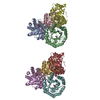
| ||||||||||||||||||||||||||||||||
|---|---|---|---|---|---|---|---|---|---|---|---|---|---|---|---|---|---|---|---|---|---|---|---|---|---|---|---|---|---|---|---|---|---|
| 1 | 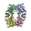
| ||||||||||||||||||||||||||||||||
| 2 | 
| ||||||||||||||||||||||||||||||||
| Unit cell |
| ||||||||||||||||||||||||||||||||
| Noncrystallographic symmetry (NCS) | NCS oper:
|
-Components
| #1: Protein | Mass: 23723.691 Da / Num. of mol.: 8 Source method: isolated from a genetically manipulated source Details: 2-CARBOXYETHYLPHOSPHONIC ACID IN THE ACTIVE SITE / Source: (gene. exp.) PYROCOCCUS WOESEI (archaea) / Production host:  References: UniProt: P95583, UniProt: P62003*PLUS, triose-phosphate isomerase #2: Chemical | ChemComp-3PP /   Mass: 154.058 Da / Num. of mol.: 8 / Source method: obtained synthetically / Formula: C3H7O5P Mass: 154.058 Da / Num. of mol.: 8 / Source method: obtained synthetically / Formula: C3H7O5P#3: Water | ChemComp-HOH / |  Mass: 18.015 Da / Num. of mol.: 346 / Source method: isolated from a natural source / Formula: H2O Mass: 18.015 Da / Num. of mol.: 346 / Source method: isolated from a natural source / Formula: H2OCompound details | CONVERTS D-GLYCERALDEHYDE 3-PHOSPHATE TO DIHYDROXY-ACETONE PHOSPHATE. PLAYS AN IMPORTANT ROLE IN ...CONVERTS D-GLYCERALDE | Sequence details | THE C-TERMINAL 15 RESIDUES LISTED IN THE SWISS-PROT ENTRY ARE INCORRECT. THE SWISS-PROT ENTRY WILL ...THE C-TERMINAL 15 RESIDUES LISTED IN THE SWISS-PROT ENTRY ARE INCORRECT. THE SWISS-PROT ENTRY WILL BE UPDATED SOON. | |
|---|
-Experimental details
-Experiment
| Experiment | Method: X-RAY DIFFRACTION / Number of used crystals: 2 |
|---|
- Sample preparation
| Crystal | Density Matthews: 2.7 Å3/Da / Density % sol: 55 % Description: MAD DATA WERE USED TO DETERMINE THE STRUCTURE, CROSS- CRYSTAL AVERAGING AND PHASE EXTENSION USING IN-HOUSE DATA WERE USED IN REFINEMENT OF THE MODEL. | |||||||||||||||||||||||||
|---|---|---|---|---|---|---|---|---|---|---|---|---|---|---|---|---|---|---|---|---|---|---|---|---|---|---|
| Crystal grow | pH: 4.6 / Details: 0.1M SODIUM ACETATE PH 4.0 AND 7% PEG 4000 | |||||||||||||||||||||||||
| Crystal grow | *PLUS pH: 4 / Method: vapor diffusion, hanging drop / Details: Bell, G.S., (1998) Acta crystallog., D54, 1419. | |||||||||||||||||||||||||
| Components of the solutions | *PLUS
|
-Data collection
| Diffraction | Mean temperature: 100 K | ||||||||||||
|---|---|---|---|---|---|---|---|---|---|---|---|---|---|
| Diffraction source | Source: SYNCHROTRON / Site: EMBL/DESY, HAMBURG  / Beamline: BW7A / Wavelength: 0.9795,0.9796,0.9537 / Beamline: BW7A / Wavelength: 0.9795,0.9796,0.9537 | ||||||||||||
| Radiation | Protocol: MAD / Monochromatic (M) / Laue (L): M / Scattering type: x-ray | ||||||||||||
| Radiation wavelength |
| ||||||||||||
| Reflection | Resolution: 3→30 Å / Num. obs: 25844 / % possible obs: 90.1 % / Observed criterion σ(I): 2.5 / Redundancy: 15 % / Biso Wilson estimate: 28 Å2 / Rmerge(I) obs: 0.076 | ||||||||||||
| Reflection shell | Resolution: 3→3.2 Å / Rmerge(I) obs: 0.112 / % possible all: 89.7 | ||||||||||||
| Reflection | *PLUS Num. measured all: 386571 | ||||||||||||
| Reflection shell | *PLUS % possible obs: 89.7 % |
- Processing
| Software |
| ||||||||||||||||||||||||||||||||||||||||||||||||||||||||||||
|---|---|---|---|---|---|---|---|---|---|---|---|---|---|---|---|---|---|---|---|---|---|---|---|---|---|---|---|---|---|---|---|---|---|---|---|---|---|---|---|---|---|---|---|---|---|---|---|---|---|---|---|---|---|---|---|---|---|---|---|---|---|
| Refinement | Method to determine structure: MAD / Resolution: 2.7→6 Å / Cross valid method: THROUGHOUT / σ(F): 0 / Details: NCS RESTRAINTS WERE USED THROUGHOUT REFINEMENT
| ||||||||||||||||||||||||||||||||||||||||||||||||||||||||||||
| Displacement parameters | Biso mean: 26 Å2 | ||||||||||||||||||||||||||||||||||||||||||||||||||||||||||||
| Refinement step | Cycle: LAST / Resolution: 2.7→6 Å
| ||||||||||||||||||||||||||||||||||||||||||||||||||||||||||||
| Refine LS restraints |
| ||||||||||||||||||||||||||||||||||||||||||||||||||||||||||||
| LS refinement shell | Resolution: 2.7→2.8 Å | ||||||||||||||||||||||||||||||||||||||||||||||||||||||||||||
| Software | *PLUS Name: CNS / Version: 0.9 / Classification: refinement | ||||||||||||||||||||||||||||||||||||||||||||||||||||||||||||
| Refinement | *PLUS Lowest resolution: 30 Å | ||||||||||||||||||||||||||||||||||||||||||||||||||||||||||||
| Solvent computation | *PLUS | ||||||||||||||||||||||||||||||||||||||||||||||||||||||||||||
| Displacement parameters | *PLUS |
