 Movie
Movie Controller

[English] 日本語
 Yorodumi
- PDB-1gx1: Structure of 2C-Methyl-D-erythritol-2,4-cyclodiphosphate Synthase -
+ Open data

- Basic information
| Entry | Database: PDB / ID: 1gx1 | ||||||
|---|---|---|---|---|---|---|---|
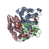
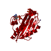
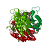
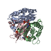
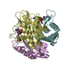
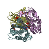
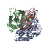
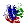
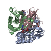
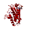
| Title | Structure of 2C-Methyl-D-erythritol-2,4-cyclodiphosphate Synthase | ||||||
 Components Components | 2-C-METHYL-D-ERYTHRITOL 2,4-CYCLODIPHOSPHATE SYNTHASE | ||||||
 Keywords Keywords | SYNTHASE / ISOPRENOID / LYASE / ISOPRENE BIOSYNTHESIS | ||||||
| Function / homology |  Function and homology information Function and homology information2-C-methyl-D-erythritol 2,4-cyclodiphosphate synthase / 2-C-methyl-D-erythritol 2,4-cyclodiphosphate synthase activity / isopentenyl diphosphate biosynthetic process, methylerythritol 4-phosphate pathway / terpenoid biosynthetic process / ubiquinone biosynthetic process / manganese ion binding / zinc ion binding / metal ion binding / identical protein binding Similarity search - Function | ||||||
| Biological species |  | ||||||
| Method |  X-RAY DIFFRACTION / SYNCHROTRON / MAD / Resolution: 1.8 Å X-RAY DIFFRACTION / SYNCHROTRON / MAD / Resolution: 1.8 Å | ||||||
 Authors Authors | Kemp, L.E. / Bond, C.S. / Hunter, W.N. | ||||||
 Citation Citation | Journal: Proc.Natl.Acad.Sci.USA / Year: 2002 Title: Structure of 2C-Methyl-D-Erythritol 2,4-Cyclodiphosphate Synthase: An Essential Enzyme for Isoprenoid Biosynthesis and Target for Antimicrobial Drug Development Authors: Kemp, L.E. / Bond, C.S. / Hunter, W.N. | ||||||
| History |
|
- Structure visualization
| Structure viewer | Molecule: MolmilJmol/JSmol |
|---|
- Downloads & links
-Download
| PDBx/mmCIF format | 1gx1.cif.gz | 111.8 KB | Display | PDBx/mmCIF format |
|---|---|---|---|---|
| PDB format | pdb1gx1.ent.gz | 86.5 KB | Display | PDB format |
| PDBx/mmJSON format | 1gx1.json.gz | Tree view | PDBx/mmJSON format | |
| Others |  Other downloads Other downloads |
-Validation report
| Arichive directory | https://data.pdbj.org/pub/pdb/validation_reports/gx/1gx1ftp://data.pdbj.org/pub/pdb/validation_reports/gx/1gx1 | HTTPS FTP |
|---|
-Related structure data
| Related structure data | |
|---|---|
| Similar structure data |

-Links
 PDBj
PDBj- Assembly
| Deposited unit | 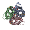
| ||||||||
|---|---|---|---|---|---|---|---|---|---|
| 1 |
| ||||||||
| Unit cell |
| ||||||||
| Noncrystallographic symmetry (NCS) | NCS oper: (Code: given Matrix: (-0.483668, 0.118894, -0.867139), Vector: |
-Components
-Protein , 1 types, 3 molecules ABC
| #1: Protein | Mass: 17237.229 Da / Num. of mol.: 3 Source method: isolated from a genetically manipulated source Source: (gene. exp.) References: UniProt: P36663, UniProt: P62617*PLUS, 2-C-methyl-D-erythritol 2,4-cyclodiphosphate synthase |
|---|
-Non-polymers , 5 types, 309 molecules 


| #2: Chemical |  Mass: 65.409 Da / Num. of mol.: 3 / Source method: obtained synthetically / Formula: Zn Mass: 65.409 Da / Num. of mol.: 3 / Source method: obtained synthetically / Formula: Zn#3: Chemical |  Mass: 403.176 Da / Num. of mol.: 3 / Source method: obtained synthetically / Formula: C9H15N3O11P2 Mass: 403.176 Da / Num. of mol.: 3 / Source method: obtained synthetically / Formula: C9H15N3O11P2#4: Chemical |  Mass: 54.938 Da / Num. of mol.: 3 / Source method: obtained synthetically / Formula: Mn Mass: 54.938 Da / Num. of mol.: 3 / Source method: obtained synthetically / Formula: Mn#5: Chemical | ChemComp-SO4 / |  Mass: 96.063 Da / Num. of mol.: 1 / Source method: obtained synthetically / Formula: SO4 Mass: 96.063 Da / Num. of mol.: 1 / Source method: obtained synthetically / Formula: SO4#6: Water | ChemComp-HOH / | Mass: 18.015 Da / Num. of mol.: 299 / Source method: isolated from a natural source / Formula: H2O |
|---|
-Details
| Has protein modification | Y |
|---|
-Experimental details
-Experiment
| Experiment | Method: X-RAY DIFFRACTION / Number of used crystals: 1 |
|---|
- Sample preparation
| Crystal | Density Matthews: 2.4 Å3/Da / Density % sol: 48.3 % | |||||||||||||||||||||||||||||||||||||||||||||||||
|---|---|---|---|---|---|---|---|---|---|---|---|---|---|---|---|---|---|---|---|---|---|---|---|---|---|---|---|---|---|---|---|---|---|---|---|---|---|---|---|---|---|---|---|---|---|---|---|---|---|---|
| Crystal grow | pH: 4.6 Details: 10-20% PEG 2000 MONOETHYL ETHER, 0.1M AMMONIUM SULPHATE, 0.1M SODIUM ACETATE, pH 4.60 | |||||||||||||||||||||||||||||||||||||||||||||||||
| Crystal grow | *PLUS pH: 7.7 / Method: vapor diffusion, hanging drop | |||||||||||||||||||||||||||||||||||||||||||||||||
| Components of the solutions | *PLUS
|
-Data collection
| Diffraction | Mean temperature: 100 K | ||||||||||||
|---|---|---|---|---|---|---|---|---|---|---|---|---|---|
| Diffraction source | Source: SYNCHROTRON / Site: ESRF  / Beamline: ID29 / Wavelength: 0.9184,0.9786,0.9788 / Beamline: ID29 / Wavelength: 0.9184,0.9786,0.9788 | ||||||||||||
| Detector | Type: MARRESEARCH / Detector: CCD / Date: Mar 15, 2001 / Details: MIRRORS | ||||||||||||
| Radiation | Monochromator: SI / Protocol: MAD / Monochromatic (M) / Laue (L): M / Scattering type: x-ray | ||||||||||||
| Radiation wavelength |
| ||||||||||||
| Reflection | Resolution: 1.8→30 Å / Num. obs: 91076 / % possible obs: 92.8 % / Observed criterion σ(I): 2 / Redundancy: 2.1 % / Rmerge(I) obs: 0.06 / Net I/σ(I): 12.6 | ||||||||||||
| Reflection shell | Resolution: 1.8→1.86 Å / Rmerge(I) obs: 0.279 / Mean I/σ(I) obs: 1.7 / % possible all: 73.8 | ||||||||||||
| Reflection | *PLUS Lowest resolution: 30 Å / Num. obs: 50289 / % possible obs: 97.4 % / Num. measured all: 193317 / Rmerge(I) obs: 0.067 | ||||||||||||
| Reflection shell | *PLUS % possible obs: 89.5 % / Rmerge(I) obs: 0.32 / Mean I/σ(I) obs: 2 |
- Processing
| Software |
| ||||||||||||||||||||
|---|---|---|---|---|---|---|---|---|---|---|---|---|---|---|---|---|---|---|---|---|---|
| Refinement | Method to determine structure: MAD / Resolution: 1.8→20 Å / SU B: 2.473 / SU ML: 0.077 / Cross valid method: THROUGHOUT / ESU R Free: 0.113 Details: THERE ARE RESIDUES WHOSE SIDECHAINS COULD NOT BE FITTED. THESE ARE INCLUDED IN THE PDB FILE WITH ZERO OCCUPANCY
| ||||||||||||||||||||
| Refinement step | Cycle: LAST / Resolution: 1.8→20 Å
| ||||||||||||||||||||
| Refinement | *PLUS Highest resolution: 1.8 Å / Lowest resolution: 20 Å / Rfactor obs: 0.18 / Rfactor Rfree: 0.213 / Rfactor Rwork: 0.18 | ||||||||||||||||||||
| Solvent computation | *PLUS | ||||||||||||||||||||
| Displacement parameters | *PLUS | ||||||||||||||||||||
| Refine LS restraints | *PLUS
|
