 Movie
Movie Controller

[English] 日本語
 Yorodumi
- PDB-1g4a: CRYSTAL STRUCTURES OF THE HSLVU PEPTIDASE-ATPASE COMPLEX REVEAL A... -
+ Open data

- Basic information
| Entry | Database: PDB / ID: 1g4a | ||||||
|---|---|---|---|---|---|---|---|
| Title | CRYSTAL STRUCTURES OF THE HSLVU PEPTIDASE-ATPASE COMPLEX REVEAL AN ATP-DEPENDENT PROTEOLYSIS MECHANISM | ||||||
 Components Components |
| ||||||
 Keywords Keywords | CHAPERONE/HYDROLASE / HSLVU / PEPTIDASE-ATPASE COMPLEX / CHAPERONE-HYDROLASE COMPLEX | ||||||
| Function / homology |  Function and homology information Function and homology informationprotein denaturation / HslU-HslV peptidase / HslUV protease complex / proteasome-activating activity / proteasome core complex / protein unfolding / threonine-type endopeptidase activity / : / peptidase activity / cellular response to heat ...protein denaturation / HslU-HslV peptidase / HslUV protease complex / proteasome-activating activity / proteasome core complex / protein unfolding / threonine-type endopeptidase activity / : / peptidase activity / cellular response to heat / response to heat / protein domain specific binding / magnesium ion binding / ATP hydrolysis activity / proteolysis / ATP binding / membrane / identical protein binding / cytosol / cytoplasm Similarity search - Function | ||||||
| Biological species |  | ||||||
| Method |  X-RAY DIFFRACTION / SYNCHROTRON / Resolution: 3 Å X-RAY DIFFRACTION / SYNCHROTRON / Resolution: 3 Å | ||||||
 Authors Authors | Wang, J. / Song, J.J. / Franklin, M.C. / Kamtekar, S. / Im, Y.J. / Rho, S.H. / Seong, I.S. / Lee, C.S. / Chung, C.H. / Eom, S.H. | ||||||
 Citation Citation | Journal: Structure / Year: 2001 Title: Crystal structures of the HslVU peptidase-ATPase complex reveal an ATP-dependent proteolysis mechanism. Authors: Wang, J. / Song, J.J. / Franklin, M.C. / Kamtekar, S. / Im, Y.J. / Rho, S.H. / Seong, I.S. / Lee, C.S. / Chung, C.H. / Eom, S.H. #1: Journal: J.Biol.Chem. / Year: 1996Title: Purification and Characterization of the Heat Shock Proteins HslV and HslU that form a New ATP-Dependent Protease in Escherichia Coli Authors: Yoo, S.J. / Seol, J.H. / Shin, D.H. / Rohrwild, M. / Kang, M.S. / Tanaka, K. / Goldberg, A.L. / Chung, C.H. #2: Journal: Nature / Year: 2000Title: The Structuers of HslU and the ATP-Dependent Protease HslU-HslV. Authors: Bochtler, M. / Hartmann, C. / Song, H.K. / Bourenkov, G.P. / Bartunik, H.D. / Huber, R. | ||||||
| History |
|
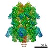
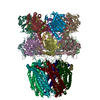
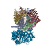
- Structure visualization
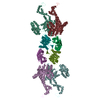
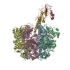
| Structure viewer | Molecule: MolmilJmol/JSmol |
|---|
- Downloads & links
-Download
| PDBx/mmCIF format | 1g4a.cif.gz | 279.9 KB | Display | PDBx/mmCIF format |
|---|---|---|---|---|
| PDB format | pdb1g4a.ent.gz | 222.8 KB | Display | PDB format |
| PDBx/mmJSON format | 1g4a.json.gz | Tree view | PDBx/mmJSON format | |
| Others |  Other downloads Other downloads |
-Validation report
| Arichive directory | https://data.pdbj.org/pub/pdb/validation_reports/g4/1g4aftp://data.pdbj.org/pub/pdb/validation_reports/g4/1g4a | HTTPS FTP |
|---|
-Related structure data

-Links
 PDBj
PDBj


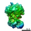
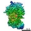
- Assembly
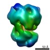
| Deposited unit | 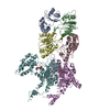
| ||||||||
|---|---|---|---|---|---|---|---|---|---|
| 1 | 
| ||||||||
| Unit cell |
|
-Components
| #1: Protein | Mass: 49659.703 Da / Num. of mol.: 2 Source method: isolated from a genetically manipulated source Source: (gene. exp.) #2: Protein | Mass: 18986.641 Da / Num. of mol.: 4 Source method: isolated from a genetically manipulated source Source: (gene. exp.) #3: Chemical |   Mass: 411.202 Da / Num. of mol.: 2 / Source method: obtained synthetically / Formula: C10H15N5O9P2 Mass: 411.202 Da / Num. of mol.: 2 / Source method: obtained synthetically / Formula: C10H15N5O9P2 |
|---|
-Experimental details
-Experiment
| Experiment | Method: X-RAY DIFFRACTION / Number of used crystals: 1 |
|---|
- Sample preparation
| Crystal | Density Matthews: 3.84 Å3/Da / Density % sol: 67.95 % | ||||||||||||||||||||||||||||||||||||||||||||||||||||||||||||||||||
|---|---|---|---|---|---|---|---|---|---|---|---|---|---|---|---|---|---|---|---|---|---|---|---|---|---|---|---|---|---|---|---|---|---|---|---|---|---|---|---|---|---|---|---|---|---|---|---|---|---|---|---|---|---|---|---|---|---|---|---|---|---|---|---|---|---|---|---|
| Crystal grow | Method: vapor diffusion / Details: dADP-HslU-HslV, VAPOR DIFFUSION | ||||||||||||||||||||||||||||||||||||||||||||||||||||||||||||||||||
| Crystal grow | *PLUS pH: 7.8 / Method: vapor diffusion, hanging drop | ||||||||||||||||||||||||||||||||||||||||||||||||||||||||||||||||||
| Components of the solutions | *PLUS
|
-Data collection
| Diffraction source | Source: SYNCHROTRON / Site: ALS  / Beamline: 5.0.3 / Beamline: 5.0.3 |
|---|---|
| Radiation | Protocol: SINGLE WAVELENGTH / Monochromatic (M) / Laue (L): M / Scattering type: x-ray |
| Radiation wavelength | Relative weight: 1 |
| Reflection | *PLUS Highest resolution: 3 Å / Lowest resolution: 85 Å / Num. obs: 46551 / % possible obs: 86 % / Rmerge(I) obs: 0.131 |
- Processing
| Software |
| ||||||||||||||||||||||||||||||||||||||||||||||||||||||||||||
|---|---|---|---|---|---|---|---|---|---|---|---|---|---|---|---|---|---|---|---|---|---|---|---|---|---|---|---|---|---|---|---|---|---|---|---|---|---|---|---|---|---|---|---|---|---|---|---|---|---|---|---|---|---|---|---|---|---|---|---|---|---|
| Refinement | Resolution: 3→85 Å / Rfactor Rfree error: 0.006 / Data cutoff high absF: 145156.31 / Data cutoff low absF: 0 / Isotropic thermal model: RESTRAINED / Cross valid method: THROUGHOUT / σ(F): 0
| ||||||||||||||||||||||||||||||||||||||||||||||||||||||||||||
| Solvent computation | Solvent model: FLAT MODEL / Bsol: 31.98 Å2 / ksol: 0.295 e/Å3 | ||||||||||||||||||||||||||||||||||||||||||||||||||||||||||||
| Displacement parameters | Biso mean: 52.1 Å2
| ||||||||||||||||||||||||||||||||||||||||||||||||||||||||||||
| Refinement step | Cycle: LAST / Resolution: 3→85 Å
| ||||||||||||||||||||||||||||||||||||||||||||||||||||||||||||
| Refine LS restraints |
| ||||||||||||||||||||||||||||||||||||||||||||||||||||||||||||
| LS refinement shell | Resolution: 3→3.19 Å / Rfactor Rfree error: 0.016 / Total num. of bins used: 6
| ||||||||||||||||||||||||||||||||||||||||||||||||||||||||||||
| Xplor file |
| ||||||||||||||||||||||||||||||||||||||||||||||||||||||||||||
| Software | *PLUS Name: CNS / Version: 1 / Classification: refinement | ||||||||||||||||||||||||||||||||||||||||||||||||||||||||||||
| Refinement | *PLUS σ(F): 0 / % reflection Rfree: 10 % | ||||||||||||||||||||||||||||||||||||||||||||||||||||||||||||
| Solvent computation | *PLUS | ||||||||||||||||||||||||||||||||||||||||||||||||||||||||||||
| Displacement parameters | *PLUS Biso mean: 52.1 Å2 | ||||||||||||||||||||||||||||||||||||||||||||||||||||||||||||
| Refine LS restraints | *PLUS
| ||||||||||||||||||||||||||||||||||||||||||||||||||||||||||||
| LS refinement shell | *PLUS % reflection Rfree: 10.8 % |
