 Movie
Movie Controller

[English] 日本語
 Yorodumi
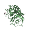
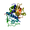
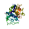
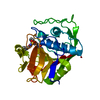
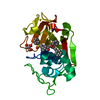
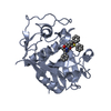
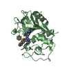
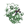
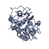
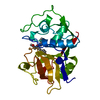
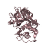
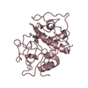
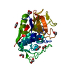
- PDB-1f2b: CRYSTAL STRUCTURE ANALYSIS OF CRUZAIN BOUND TO VINYL SULFONE DERI... -
+ Open data

- Basic information
| Entry | Database: PDB / ID: 1f2b | ||||||
|---|---|---|---|---|---|---|---|
| Title | CRYSTAL STRUCTURE ANALYSIS OF CRUZAIN BOUND TO VINYL SULFONE DERIVED INHIBITOR (III) | ||||||
 Components Components | CRUZAIN | ||||||
 Keywords Keywords | HYDROLASE / cysteine protease / covalent inhibitor / vinyl sulfone-derived / P1' pocket | ||||||
| Function / homology |  Function and homology information Function and homology information | ||||||
| Biological species |  | ||||||
| Method |  X-RAY DIFFRACTION / Resolution: 1.8 Å X-RAY DIFFRACTION / Resolution: 1.8 Å | ||||||
 Authors Authors | Brinen, L.S. / Hansell, E. / Roush, W.R. / McKerrow, J.H. / Fletterick, R.J. | ||||||
 Citation Citation | Journal: Structure Fold.Des. / Year: 2000 Title: A target within the target: probing cruzain's P1' site to define structural determinants for the Chagas' disease protease. Authors: Brinen, L.S. / Hansell, E. / Cheng, J. / Roush, W.R. / McKerrow, J.H. / Fletterick, R.J. #1: Journal: J.Mol.Biol. / Year: 1995Title: The Crystal Structure of Cruzain: a Therapeutic Target for Chagas' Disease Authors: McGrath, M.E. / Eakin, A.E. / Engel, J.C. / McKerrow, J.H. / Craik, C.S. / Fletterick, R.J. | ||||||
| History |
|
- Structure visualization
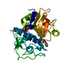
| Structure viewer | Molecule: MolmilJmol/JSmol |
|---|
- Downloads & links
-Download
| PDBx/mmCIF format | 1f2b.cif.gz | 55.2 KB | Display | PDBx/mmCIF format |
|---|---|---|---|---|
| PDB format | pdb1f2b.ent.gz | 38.7 KB | Display | PDB format |
| PDBx/mmJSON format | 1f2b.json.gz | Tree view | PDBx/mmJSON format | |
| Others |  Other downloads Other downloads |
-Validation report
| Arichive directory | https://data.pdbj.org/pub/pdb/validation_reports/f2/1f2bftp://data.pdbj.org/pub/pdb/validation_reports/f2/1f2b | HTTPS FTP |
|---|
-Related structure data


-Links
 PDBj
PDBj
- Assembly
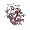
| Deposited unit | 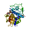
| ||||||||
|---|---|---|---|---|---|---|---|---|---|
| 1 |
| ||||||||
| Unit cell |
|
-Components
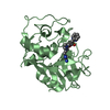
| #1: Protein | Mass: 22715.133 Da / Num. of mol.: 1 / Fragment: CATALYTIC DOMAIN Source method: isolated from a genetically manipulated source Source: (gene. exp.)  References: UniProt: P25779, Hydrolases; Acting on peptide bonds (peptidases); Cysteine endopeptidases |
|---|---|
| #2: Chemical | ChemComp-VS3 / 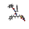  Mass: 645.722 Da / Num. of mol.: 1 / Source method: obtained synthetically / Formula: C34H35N3O8S Mass: 645.722 Da / Num. of mol.: 1 / Source method: obtained synthetically / Formula: C34H35N3O8S |
| #3: Water | ChemComp-HOH /  Mass: 18.015 Da / Num. of mol.: 66 / Source method: isolated from a natural source / Formula: H2O Mass: 18.015 Da / Num. of mol.: 66 / Source method: isolated from a natural source / Formula: H2O |
| Has protein modification | Y |
-Experimental details
-Experiment
| Experiment | Method: X-RAY DIFFRACTION / Number of used crystals: 1 |
|---|
- Sample preparation
| Crystal | Density Matthews: 2.09 Å3/Da / Density % sol: 41.09 % | ||||||||||||||||||||
|---|---|---|---|---|---|---|---|---|---|---|---|---|---|---|---|---|---|---|---|---|---|
| Crystal grow | Temperature: 291 K / Method: vapor diffusion, hanging drop / pH: 6.5 Details: Sodium citrate; micro seeding, pH 6.5, VAPOR DIFFUSION, HANGING DROP, temperature 291K | ||||||||||||||||||||
| Crystal grow | *PLUS Temperature: 18 ℃ / pH: 5.8 | ||||||||||||||||||||
| Components of the solutions | *PLUS
|
-Data collection
| Diffraction | Mean temperature: 298 K |
|---|---|
| Diffraction source | Source: ROTATING ANODE / Type: RIGAKU RU300 / Wavelength: 1.5418 |
| Detector | Type: MARRESEARCH / Detector: IMAGE PLATE / Date: Sep 15, 1998 |
| Radiation | Protocol: SINGLE WAVELENGTH / Monochromatic (M) / Laue (L): M / Scattering type: x-ray |
| Radiation wavelength | Wavelength: 1.5418 Å / Relative weight: 1 |
| Reflection | Resolution: 1.7→30 Å / Num. all: 17495 / Num. obs: 15513 / % possible obs: 88.8 % / Observed criterion σ(F): 0 / Observed criterion σ(I): 0 / Redundancy: 7.8 % / Biso Wilson estimate: 12.6 Å2 / Rmerge(I) obs: 0.08 / Net I/σ(I): 12.4 |
| Reflection shell | Resolution: 1.8→1.85 Å / Redundancy: 1.7 % / Rmerge(I) obs: 0.22 / % possible all: 74.4 |
| Reflection shell | *PLUS % possible obs: 74.4 % |
- Processing
| Software |
| |||||||||||||||||||||||||
|---|---|---|---|---|---|---|---|---|---|---|---|---|---|---|---|---|---|---|---|---|---|---|---|---|---|---|
| Refinement | Resolution: 1.8→30 Å / σ(F): 2 / σ(I): 2 / Stereochemistry target values: Engh & Huber
| |||||||||||||||||||||||||
| Refinement step | Cycle: LAST / Resolution: 1.8→30 Å
| |||||||||||||||||||||||||
| Refine LS restraints |
| |||||||||||||||||||||||||
| Software | *PLUS Name: X-PLOR / Version: 3.843 / Classification: refinement | |||||||||||||||||||||||||
| Refine LS restraints | *PLUS
|
