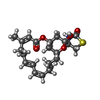
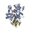
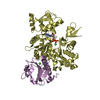
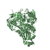
Entry Database : PDB / ID : 1esvTitle COMPLEX BETWEEN LATRUNCULIN A:RABBIT MUSCLE ALPHA ACTIN:HUMAN GELSOLIN DOMAIN 1 Keywords / / / / / Function / homology Function Domain/homology Component
/ / / / / / / / / / / / / / / / / / / / / / / / / / / / / / / / / / / / / / / / / / / / / / / / / / / / / / / / / / / / / / / / / / / / / / / / / / / / / / / / / / / / / / / / / / / / / / / / / / / / / / / / / / / / / / / / / / / / / / / / / / / / / Biological species Homo sapiens (human)Oryctolagus cuniculus (rabbit)Method / / Resolution : 2 Å Authors Morton, W.M. / Ayscough, K.A. / McLaughlin, P.J. Journal : Nat.Cell Biol. / Year : 2000Title : Latrunculin alters the actin-monomer subunit interface to prevent polymerization.Authors : Morton, W.M. / Ayscough, K.R. / McLaughlin, P.J. History Deposition Apr 11, 2000 Deposition site / Processing site Revision 1.0 Jul 19, 2000 Provider / Type Revision 1.1 Apr 27, 2008 Group Revision 1.2 Jul 13, 2011 Group Revision 1.3 Nov 3, 2021 Group / Derived calculationsCategory database_2 / pdbx_struct_conn_angle ... database_2 / pdbx_struct_conn_angle / struct_conn / struct_ref_seq_dif / struct_site Item _database_2.pdbx_DOI / _database_2.pdbx_database_accession ... _database_2.pdbx_DOI / _database_2.pdbx_database_accession / _pdbx_struct_conn_angle.ptnr1_auth_asym_id / _pdbx_struct_conn_angle.ptnr1_auth_comp_id / _pdbx_struct_conn_angle.ptnr1_auth_seq_id / _pdbx_struct_conn_angle.ptnr1_label_asym_id / _pdbx_struct_conn_angle.ptnr1_label_atom_id / _pdbx_struct_conn_angle.ptnr1_label_comp_id / _pdbx_struct_conn_angle.ptnr1_label_seq_id / _pdbx_struct_conn_angle.ptnr2_auth_asym_id / _pdbx_struct_conn_angle.ptnr2_auth_seq_id / _pdbx_struct_conn_angle.ptnr2_label_asym_id / _pdbx_struct_conn_angle.ptnr3_auth_asym_id / _pdbx_struct_conn_angle.ptnr3_auth_comp_id / _pdbx_struct_conn_angle.ptnr3_auth_seq_id / _pdbx_struct_conn_angle.ptnr3_label_asym_id / _pdbx_struct_conn_angle.ptnr3_label_atom_id / _pdbx_struct_conn_angle.ptnr3_label_comp_id / _pdbx_struct_conn_angle.ptnr3_label_seq_id / _pdbx_struct_conn_angle.value / _struct_conn.pdbx_dist_value / _struct_conn.pdbx_leaving_atom_flag / _struct_conn.ptnr1_auth_asym_id / _struct_conn.ptnr1_auth_comp_id / _struct_conn.ptnr1_auth_seq_id / _struct_conn.ptnr1_label_asym_id / _struct_conn.ptnr1_label_atom_id / _struct_conn.ptnr1_label_comp_id / _struct_conn.ptnr1_label_seq_id / _struct_conn.ptnr2_auth_asym_id / _struct_conn.ptnr2_auth_comp_id / _struct_conn.ptnr2_auth_seq_id / _struct_conn.ptnr2_label_asym_id / _struct_conn.ptnr2_label_atom_id / _struct_conn.ptnr2_label_comp_id / _struct_conn.ptnr2_label_seq_id / _struct_ref_seq_dif.details / _struct_site.pdbx_auth_asym_id / _struct_site.pdbx_auth_comp_id / _struct_site.pdbx_auth_seq_id
Show all Show less
 Yorodumi
 Open data
 Basic information
 Components
 Keywords
 Function and homology information
 Homo sapiens (human)
 X-RAY DIFFRACTION /
  SYNCHROTRON / Resolution: 2 Å
  Authors
 Citation
 Journal: Nat.Cell Biol. / Year: 2000
 Structure visualization
 Molmil
 Jmol/JSmol
 Downloads & links
 Download
 1esv.cif.gz
 PDBx/mmCIF format
 pdb1esv.ent.gz
 PDB format
 1esv.json.gz
 PDBx/mmJSON format
 Other downloads
 1esv_validation.pdf.gz
 wwPDB validaton report
 1esv_full_validation.pdf.gz
 1esv_validation.xml.gz
 1esv_validation.cif.gz
 https://data.pdbj.org/pub/pdb/validation_reports/es/1esv
 ftp://data.pdbj.org/pub/pdb/validation_reports/es/1esv
 Links
Links Assembly
 Components
 Homo sapiens (human) / Cell: PLASMA / Plasmid: PMW172 / Production host:




 X-RAY DIFFRACTION / Number of used crystals: 1
  Sample preparation
 SYNCHROTRON / Site:
  SRS
SRS  / Beamline: PX9.6 / Wavelength: 0.87
  Processing
 Movie
Movie Controller


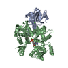
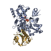

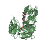
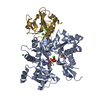
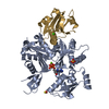
 PDBj
PDBj


