 Movie
Movie Controller

[English] 日本語
 Yorodumi
- PDB-1dfo: CRYSTAL STRUCTURE AT 2.4 ANGSTROM RESOLUTION OF E. COLI SERINE HY... -
+ Open data

- Basic information
| Entry | Database: PDB / ID: 1dfo | ||||||
|---|---|---|---|---|---|---|---|
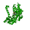
| Title | CRYSTAL STRUCTURE AT 2.4 ANGSTROM RESOLUTION OF E. COLI SERINE HYDROXYMETHYLTRANSFERASE IN COMPLEX WITH GLYCINE AND 5-FORMYL TETRAHYDROFOLATE | ||||||
 Components Components | SERINE HYDROXYMETHYLTRANSFERASE | ||||||
 Keywords Keywords | TRANSFERASE / ALPHA PLP ASPARTATE / AMINO TRANSFERASE / (AAT)-LIKE FOLD | ||||||
| Function / homology |  Function and homology information Function and homology informationglycine catabolic process / L-allo-threonine aldolase activity / L-serine biosynthetic process / glycine hydroxymethyltransferase / glycine hydroxymethyltransferase activity / glycine biosynthetic process from serine / serine binding / L-serine catabolic process / tetrahydrofolate metabolic process / tetrahydrofolate interconversion ...glycine catabolic process / L-allo-threonine aldolase activity / L-serine biosynthetic process / glycine hydroxymethyltransferase / glycine hydroxymethyltransferase activity / glycine biosynthetic process from serine / serine binding / L-serine catabolic process / tetrahydrofolate metabolic process / tetrahydrofolate interconversion / cobalt ion binding / folic acid metabolic process / pyridoxal phosphate binding / protein homodimerization activity / zinc ion binding / identical protein binding / membrane / cytoplasm / cytosol Similarity search - Function | ||||||
| Biological species |  | ||||||
| Method |  X-RAY DIFFRACTION / Resolution: 2.4 Å X-RAY DIFFRACTION / Resolution: 2.4 Å | ||||||
 Authors Authors | Scarsdale, J.N. / Radaev, S. / Kazanina, G. / Schirch, V. / Wright, H.T. | ||||||
 Citation Citation | Journal: J.Mol.Biol. / Year: 2000 Title: Crystal structure at 2.4 A resolution of E. coli serine hydroxymethyltransferase in complex with glycine substrate and 5-formyl tetrahydrofolate. Authors: Scarsdale, J.N. / Radaev, S. / Kazanina, G. / Schirch, V. / Wright, H.T. | ||||||
| History |
|
- Structure visualization
| Structure viewer | Molecule: MolmilJmol/JSmol |
|---|
- Downloads & links
-Download
| PDBx/mmCIF format | 1dfo.cif.gz | 326.2 KB | Display | PDBx/mmCIF format |
|---|---|---|---|---|
| PDB format | pdb1dfo.ent.gz | 266.7 KB | Display | PDB format |
| PDBx/mmJSON format | 1dfo.json.gz | Tree view | PDBx/mmJSON format | |
| Others |  Other downloads Other downloads |
-Validation report
| Summary document | 1dfo_validation.pdf.gz | 2.4 MB | Display | wwPDB validaton report |
|---|---|---|---|---|
| Full document | 1dfo_full_validation.pdf.gz | 2.5 MB | Display | |
| Data in XML | 1dfo_validation.xml.gz | 65.2 KB | Display | |
| Data in CIF | 1dfo_validation.cif.gz | 86.7 KB | Display | |
| Arichive directory | https://data.pdbj.org/pub/pdb/validation_reports/df/1dfoftp://data.pdbj.org/pub/pdb/validation_reports/df/1dfo | HTTPS FTP |
-Related structure data
| Similar structure data |
|---|


-Links
 PDBj
PDBj- Assembly
| Deposited unit | 
| ||||||||
|---|---|---|---|---|---|---|---|---|---|
| 1 | 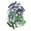
| ||||||||
| 2 | 
| ||||||||
| Unit cell |
|
-Components
| #1: Protein | Mass: 45372.477 Da / Num. of mol.: 4 Source method: isolated from a genetically manipulated source Source: (gene. exp.) Strain (production host): GS1993 (GLYA-, PHEA905, DELTA LACU169,STRA,THI, DELTAGLYA::MU, RECA-) References: UniProt: P0A825, glycine hydroxymethyltransferase #2: Chemical | ChemComp-PLG /   Mass: 306.209 Da / Num. of mol.: 4 / Source method: obtained synthetically / Formula: C10H15N2O7P Mass: 306.209 Da / Num. of mol.: 4 / Source method: obtained synthetically / Formula: C10H15N2O7P#3: Chemical | ChemComp-FFO /   Mass: 473.439 Da / Num. of mol.: 4 / Source method: obtained synthetically / Formula: C20H23N7O7 Mass: 473.439 Da / Num. of mol.: 4 / Source method: obtained synthetically / Formula: C20H23N7O7#4: Water | ChemComp-HOH / |  Mass: 18.015 Da / Num. of mol.: 388 / Source method: isolated from a natural source / Formula: H2O Mass: 18.015 Da / Num. of mol.: 388 / Source method: isolated from a natural source / Formula: H2O |
|---|
-Experimental details
-Experiment
| Experiment | Method: X-RAY DIFFRACTION / Number of used crystals: 1 |
|---|
- Sample preparation
| Crystal | Density Matthews: 3.39 Å3/Da / Density % sol: 63.68 % | ||||||||||||||||||||||||||||||
|---|---|---|---|---|---|---|---|---|---|---|---|---|---|---|---|---|---|---|---|---|---|---|---|---|---|---|---|---|---|---|---|
| Crystal grow | Temperature: 293 K / Method: vapor diffusion / pH: 7.2 Details: 2M POTASSIUM PHOSPHATE, pH 7.2, VAPOR DIFFUSION, temperature 293.0K | ||||||||||||||||||||||||||||||
| Crystal grow | *PLUS Method: vapor diffusion, hanging drop / Details: Stover, P., (1993) J.Mol.Biol., 230, 1094. | ||||||||||||||||||||||||||||||
| Components of the solutions | *PLUS
|
-Data collection
| Diffraction | Mean temperature: 293 K |
|---|---|
| Diffraction source | Source: ROTATING ANODE / Type: RIGAKU / Wavelength: 1.5418 |
| Detector | Type: RIGAKU RAXIS / Detector: IMAGE PLATE / Date: Jun 1, 1998 |
| Radiation | Protocol: SINGLE WAVELENGTH / Monochromatic (M) / Laue (L): M / Scattering type: x-ray |
| Radiation wavelength | Wavelength: 1.5418 Å / Relative weight: 1 |
| Reflection | Resolution: 2.4→91 Å / Num. all: 93964 / Num. obs: 93782 / % possible obs: 100 % / Observed criterion σ(I): -3 / Redundancy: 4.2 % / Biso Wilson estimate: 43.5 Å2 / Rmerge(I) obs: 0.23 / Net I/σ(I): 7.1 |
| Reflection shell | Resolution: 2.41→2.49 Å / Redundancy: 4 % / Rmerge(I) obs: 1 / % possible all: 100 |
| Reflection | *PLUS Num. obs: 83968 / % possible obs: 89 % / Biso Wilson estimate: 44 Å2 |
| Reflection shell | *PLUS % possible obs: 78 % / Mean I/σ(I) obs: 1.22 |
- Processing
| Software |
| ||||||||||||||||||||||||||||||||||||||||||||||||||||||||||||||||||||||||||||||||
|---|---|---|---|---|---|---|---|---|---|---|---|---|---|---|---|---|---|---|---|---|---|---|---|---|---|---|---|---|---|---|---|---|---|---|---|---|---|---|---|---|---|---|---|---|---|---|---|---|---|---|---|---|---|---|---|---|---|---|---|---|---|---|---|---|---|---|---|---|---|---|---|---|---|---|---|---|---|---|---|---|---|
| Refinement | Resolution: 2.4→20 Å / Rfactor Rfree error: 0.002 / Data cutoff high absF: 1937647.26 / Data cutoff low absF: 0 / Isotropic thermal model: RESTRAINED / Cross valid method: THROUGHOUT / σ(F): 2.2 / Stereochemistry target values: ENGH & HUBER
| ||||||||||||||||||||||||||||||||||||||||||||||||||||||||||||||||||||||||||||||||
| Solvent computation | Solvent model: FLAT MODEL / Bsol: 41.55 Å2 / ksol: 0.321 e/Å3 | ||||||||||||||||||||||||||||||||||||||||||||||||||||||||||||||||||||||||||||||||
| Displacement parameters | Biso mean: 36.2 Å2
| ||||||||||||||||||||||||||||||||||||||||||||||||||||||||||||||||||||||||||||||||
| Refine analyze |
| ||||||||||||||||||||||||||||||||||||||||||||||||||||||||||||||||||||||||||||||||
| Refinement step | Cycle: LAST / Resolution: 2.4→20 Å
| ||||||||||||||||||||||||||||||||||||||||||||||||||||||||||||||||||||||||||||||||
| Refine LS restraints |
| ||||||||||||||||||||||||||||||||||||||||||||||||||||||||||||||||||||||||||||||||
| LS refinement shell | Resolution: 2.4→2.55 Å / Rfactor Rfree error: 0.009 / Total num. of bins used: 6
| ||||||||||||||||||||||||||||||||||||||||||||||||||||||||||||||||||||||||||||||||
| Xplor file |
| ||||||||||||||||||||||||||||||||||||||||||||||||||||||||||||||||||||||||||||||||
| Software | *PLUS Name: 'CNS' / Classification: refinement | ||||||||||||||||||||||||||||||||||||||||||||||||||||||||||||||||||||||||||||||||
| Refinement | *PLUS Num. reflection obs: 73994 / Num. reflection Rfree: 8242 | ||||||||||||||||||||||||||||||||||||||||||||||||||||||||||||||||||||||||||||||||
| Solvent computation | *PLUS | ||||||||||||||||||||||||||||||||||||||||||||||||||||||||||||||||||||||||||||||||
| Displacement parameters | *PLUS | ||||||||||||||||||||||||||||||||||||||||||||||||||||||||||||||||||||||||||||||||
| Refine LS restraints | *PLUS
|
