 Movie
Movie Controller

[English] 日本語
 Yorodumi
- PDB-1d4o: CRYSTAL STRUCTURE OF TRANSHYDROGENASE DOMAIN III AT 1.2 ANGSTROMS... -
+ Open data

- Basic information
| Entry | Database: PDB / ID: 1d4o | ||||||
|---|---|---|---|---|---|---|---|
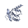
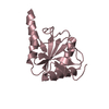
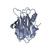
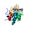
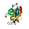
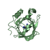
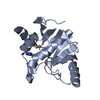
| Title | CRYSTAL STRUCTURE OF TRANSHYDROGENASE DOMAIN III AT 1.2 ANGSTROMS RESOLUTION | ||||||
 Components Components | NADP(H) TRANSHYDROGENASE | ||||||
 Keywords Keywords | OXIDOREDUCTASE / NUCLEOTIDE-BINDING FOLD / PROTEIN-NADP(H) COMPLEX / INVERTED BINDING OF NADP(H) | ||||||
| Function / homology |  Function and homology information Function and homology informationCitric acid cycle (TCA cycle) / proton-translocating NAD(P)+ transhydrogenase activity / proton-translocating NAD(P)+ transhydrogenase / NADPH regeneration / reactive oxygen species metabolic process / NADP binding / oxidoreductase activity / mitochondrial inner membrane Similarity search - Function | ||||||
| Biological species |  | ||||||
| Method |  X-RAY DIFFRACTION / SYNCHROTRON / Resolution: 1.21 Å X-RAY DIFFRACTION / SYNCHROTRON / Resolution: 1.21 Å | ||||||
 Authors Authors | Prasad, G.S. / Sridhar, V. / Yamaguchi, M. / Hatefi, Y. / Stout, C.D. | ||||||
 Citation Citation | Journal: Nat.Struct.Biol. / Year: 1999 Title: Crystal structure of transhydrogenase domain III at 1.2 A resolution. Authors: Prasad, G.S. / Sridhar, V. / Yamaguchi, M. / Hatefi, Y. / Stout, C.D. | ||||||
| History |
|
- Structure visualization
| Structure viewer | Molecule: MolmilJmol/JSmol |
|---|
- Downloads & links
-Download
| PDBx/mmCIF format | 1d4o.cif.gz | 51.7 KB | Display | PDBx/mmCIF format |
|---|---|---|---|---|
| PDB format | pdb1d4o.ent.gz | 36.6 KB | Display | PDB format |
| PDBx/mmJSON format | 1d4o.json.gz | Tree view | PDBx/mmJSON format | |
| Others |  Other downloads Other downloads |
-Validation report
| Arichive directory | https://data.pdbj.org/pub/pdb/validation_reports/d4/1d4oftp://data.pdbj.org/pub/pdb/validation_reports/d4/1d4o | HTTPS FTP |
|---|
-Related structure data
| Similar structure data |
|---|


-Links
 PDBj
PDBj

- Assembly
| Deposited unit | 
| ||||||||
|---|---|---|---|---|---|---|---|---|---|
| 1 |
| ||||||||
| Unit cell |
|
-Components
| #1: Protein | Mass: 20018.066 Da / Num. of mol.: 1 / Fragment: NADP(H) BINDING DOMAIN Source method: isolated from a genetically manipulated source Source: (gene. exp.)  References: UniProt: P11024, NAD(P)+ transhydrogenase (Si-specific) |
|---|---|
| #2: Chemical | ChemComp-NAP /   Mass: 743.405 Da / Num. of mol.: 1 / Source method: obtained synthetically / Formula: C21H28N7O17P3 Mass: 743.405 Da / Num. of mol.: 1 / Source method: obtained synthetically / Formula: C21H28N7O17P3 |
| #3: Water | ChemComp-HOH /  Mass: 18.015 Da / Num. of mol.: 185 / Source method: isolated from a natural source / Formula: H2O Mass: 18.015 Da / Num. of mol.: 185 / Source method: isolated from a natural source / Formula: H2O |
-Experimental details
-Experiment
| Experiment | Method: X-RAY DIFFRACTION / Number of used crystals: 1 |
|---|
- Sample preparation
| Crystal | Density Matthews: 2.13 Å3/Da / Density % sol: 42.15 % | |||||||||||||||||||||||||||||||||||||||||||||
|---|---|---|---|---|---|---|---|---|---|---|---|---|---|---|---|---|---|---|---|---|---|---|---|---|---|---|---|---|---|---|---|---|---|---|---|---|---|---|---|---|---|---|---|---|---|---|
| Crystal grow | Temperature: 277 K / Method: vapor diffusion, sitting drop / pH: 7.4 Details: 24% w/v MPEG 5000, 100mM sodium cacodylate and 200mM magnesium acetate., pH 7.4, VAPOR DIFFUSION, SITTING DROP | |||||||||||||||||||||||||||||||||||||||||||||
| Crystal grow | *PLUS pH: 7.8 / Method: vapor diffusion | |||||||||||||||||||||||||||||||||||||||||||||
| Components of the solutions | *PLUS
|
-Data collection
| Diffraction | Mean temperature: 100 K |
|---|---|
| Diffraction source | Source: SYNCHROTRON / Site: SSRL  / Beamline: BL7-1 / Wavelength: 1.08 / Beamline: BL7-1 / Wavelength: 1.08 |
| Detector | Type: MARRESEARCH / Detector: IMAGE PLATE / Date: Mar 21, 1999 |
| Radiation | Protocol: SINGLE WAVELENGTH / Monochromatic (M) / Laue (L): M / Scattering type: x-ray |
| Radiation wavelength | Wavelength: 1.08 Å / Relative weight: 1 |
| Reflection | Resolution: 1.21→54.1 Å / Num. all: 89452 / Num. obs: 45717 / % possible obs: 91.2 % / Observed criterion σ(I): 2.3 / Redundancy: 2 % / Biso Wilson estimate: 21.4 Å2 / Rmerge(I) obs: 0.068 / Net I/σ(I): 4 |
| Reflection shell | Resolution: 1.21→1.24 Å / Redundancy: 91.2 % / Rmerge(I) obs: 0.301 / Num. unique all: 2682 / % possible all: 72.3 |
| Reflection | *PLUS Num. measured all: 89452 |
| Reflection shell | *PLUS % possible obs: 72.3 % / Mean I/σ(I) obs: 2.3 |
- Processing
| Software |
| |||||||||||||||||||||||||
|---|---|---|---|---|---|---|---|---|---|---|---|---|---|---|---|---|---|---|---|---|---|---|---|---|---|---|
| Refinement | Resolution: 1.21→50 Å / σ(F): 0 / σ(I): 0 / Stereochemistry target values: Engh & Huber / Details: Used Shelx least squares procedure.
| |||||||||||||||||||||||||
| Refinement step | Cycle: LAST / Resolution: 1.21→50 Å
| |||||||||||||||||||||||||
| Refine LS restraints |
| |||||||||||||||||||||||||
| Software | *PLUS Name: SHELXL-97 / Classification: refinement | |||||||||||||||||||||||||
| Refinement | *PLUS σ(F): 0 / % reflection Rfree: 3 % / Rfactor Rwork: 0.167 | |||||||||||||||||||||||||
| Solvent computation | *PLUS | |||||||||||||||||||||||||
| Displacement parameters | *PLUS | |||||||||||||||||||||||||
| Refine LS restraints | *PLUS Type: s_angle_d / Dev ideal: 1.7 |
