 Movie
Movie Controller

[English] 日本語
 Yorodumi
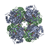
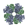
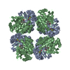
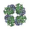
- PDB-1b4k: High resolution crystal structure of a MG2-dependent 5-aminolevul... -
+ Open data

- Basic information
| Entry | Database: PDB / ID: 1b4k | ||||||
|---|---|---|---|---|---|---|---|
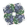
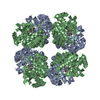
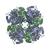
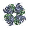
| Title | High resolution crystal structure of a MG2-dependent 5-aminolevulinic acid dehydratase | ||||||
 Components Components | PROTEIN (5-AMINOLEVULINIC ACID DEHYDRATASE) | ||||||
 Keywords Keywords | LYASE / HEME BIOSYNTHESIS / MAGNESIUM / LEVULINIC ACID | ||||||
| Function / homology |  Function and homology information Function and homology informationporphobilinogen synthase / porphobilinogen synthase activity / porphyrin-containing compound biosynthetic process / : / heme biosynthetic process / zinc ion binding / cytosol Similarity search - Function | ||||||
| Biological species |   Pseudomonas aeruginosa (bacteria) Pseudomonas aeruginosa (bacteria) | ||||||
| Method |  X-RAY DIFFRACTION / SYNCHROTRON / MOLECULAR REPLACEMENT / Resolution: 1.67 Å X-RAY DIFFRACTION / SYNCHROTRON / MOLECULAR REPLACEMENT / Resolution: 1.67 Å | ||||||
 Authors Authors | Frankenberg, N. / Jahn, D. / Heinz, D.W. | ||||||
 Citation Citation | Journal: J.Mol.Biol. / Year: 1999 Title: High resolution crystal structure of a Mg2+-dependent porphobilinogen synthase. Authors: Frankenberg, N. / Erskine, P.T. / Cooper, J.B. / Shoolingin-Jordan, P.M. / Jahn, D. / Heinz, D.W. | ||||||
| History |
|
- Structure visualization
| Structure viewer | Molecule: MolmilJmol/JSmol |
|---|
- Downloads & links
-Download
| PDBx/mmCIF format | 1b4k.cif.gz | 148.2 KB | Display | PDBx/mmCIF format |
|---|---|---|---|---|
| PDB format | pdb1b4k.ent.gz | 116.4 KB | Display | PDB format |
| PDBx/mmJSON format | 1b4k.json.gz | Tree view | PDBx/mmJSON format | |
| Others |  Other downloads Other downloads |
-Validation report
| Arichive directory | https://data.pdbj.org/pub/pdb/validation_reports/b4/1b4kftp://data.pdbj.org/pub/pdb/validation_reports/b4/1b4k | HTTPS FTP |
|---|
-Related structure data
| Related structure data |  1aw5S S: Starting model for refinement |
|---|---|
| Similar structure data |
-Links
 PDBj
PDBj
- Assembly
| Deposited unit | 
| ||||||||
|---|---|---|---|---|---|---|---|---|---|
| 1 | 
| ||||||||
| Unit cell |
| ||||||||
| Noncrystallographic symmetry (NCS) | NCS oper: (Code: given Matrix: (-0.812422, 0.582386, -0.02822), Vector: |
-Components
| #1: Protein | Mass: 37077.926 Da / Num. of mol.: 2 Source method: isolated from a genetically manipulated source Details: SCHIFF BASE LINK BETWEEN ATOM NZ OF LYS260 AND ATOM C4 OF LEVULINIC ACID Source: (gene. exp.) Pseudomonas aeruginosa (bacteria) / Strain: PAO1 / Cellular location: CYTOPLASM / Gene: HEMB / Plasmid: PGEX-6P-1 / Species (production host): Escherichia coli / Cellular location (production host): CYTOPLASM / Gene (production host): HEMB / Production host: #2: Chemical |   Mass: 96.063 Da / Num. of mol.: 2 / Source method: obtained synthetically / Formula: SO4 Mass: 96.063 Da / Num. of mol.: 2 / Source method: obtained synthetically / Formula: SO4#3: Chemical | ChemComp-MG / |   Mass: 24.305 Da / Num. of mol.: 1 / Source method: obtained synthetically / Formula: Mg Mass: 24.305 Da / Num. of mol.: 1 / Source method: obtained synthetically / Formula: Mg#4: Chemical | 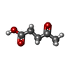  Mass: 116.115 Da / Num. of mol.: 2 / Source method: obtained synthetically / Formula: C5H8O3 Mass: 116.115 Da / Num. of mol.: 2 / Source method: obtained synthetically / Formula: C5H8O3#5: Water | ChemComp-HOH / |  Mass: 18.015 Da / Num. of mol.: 572 / Source method: isolated from a natural source / Formula: H2O Mass: 18.015 Da / Num. of mol.: 572 / Source method: isolated from a natural source / Formula: H2OHas protein modification | Y | Nonpolymer details | CARBONYL OXYGEN OF LEVULINIC ACID MISSING CARBONYL OXYGEN OF LEVULINIC ACID MISSING BECAUSE OF SCHIFF | |
|---|
-Experimental details
-Experiment
| Experiment | Method: X-RAY DIFFRACTION / Number of used crystals: 1 |
|---|
- Sample preparation
| Crystal | Density Matthews: 2.46 Å3/Da / Density % sol: 50 % | ||||||||||||||||||||||||||||||||||||||||||||||||
|---|---|---|---|---|---|---|---|---|---|---|---|---|---|---|---|---|---|---|---|---|---|---|---|---|---|---|---|---|---|---|---|---|---|---|---|---|---|---|---|---|---|---|---|---|---|---|---|---|---|
| Crystal grow | pH: 8 / Details: pH 8.0 | ||||||||||||||||||||||||||||||||||||||||||||||||
| Crystal grow | *PLUS Temperature: 20 ℃ / Method: vapor diffusion, hanging dropDetails: drop contained equal volume of protein and reservoir solution | ||||||||||||||||||||||||||||||||||||||||||||||||
| Components of the solutions | *PLUS
|
-Data collection
| Diffraction | Mean temperature: 80 K |
|---|---|
| Diffraction source | Source: SYNCHROTRON / Site: EMBL/DESY, HAMBURG  / Beamline: BW7B / Wavelength: 0.8345 / Beamline: BW7B / Wavelength: 0.8345 |
| Detector | Type: MARRESEARCH / Detector: IMAGE PLATE / Date: Jul 16, 1998 / Details: MIRRORS |
| Radiation | Protocol: SINGLE WAVELENGTH / Monochromatic (M) / Laue (L): M / Scattering type: x-ray |
| Radiation wavelength | Wavelength: 0.8345 Å / Relative weight: 1 |
| Reflection | Resolution: 1.67→51.3 Å / Num. obs: 81840 / % possible obs: 98.2 % / Redundancy: 8.8 % / Biso Wilson estimate: 20.9 Å2 / Rmerge(I) obs: 0.056 / Rsym value: 0.054 / Net I/σ(I): 9 |
| Reflection shell | Resolution: 1.67→1.76 Å / Redundancy: 4.8 % / Rmerge(I) obs: 0.301 / Mean I/σ(I) obs: 2.8 / Rsym value: 0.269 / % possible all: 94.1 |
| Reflection | *PLUS Num. measured all: 719706 / Rmerge(I) obs: 0.054 |
| Reflection shell | *PLUS % possible obs: 94.1 % / Rmerge(I) obs: 0.269 |
- Processing
| Software |
| ||||||||||||||||||||||||||||||||||||||||||||||||||||||||||||||||||||||||||||||||||||
|---|---|---|---|---|---|---|---|---|---|---|---|---|---|---|---|---|---|---|---|---|---|---|---|---|---|---|---|---|---|---|---|---|---|---|---|---|---|---|---|---|---|---|---|---|---|---|---|---|---|---|---|---|---|---|---|---|---|---|---|---|---|---|---|---|---|---|---|---|---|---|---|---|---|---|---|---|---|---|---|---|---|---|---|---|---|
| Refinement | Method to determine structure: MOLECULAR REPLACEMENT Starting model: 1AW5 Resolution: 1.67→51.3 Å / Cross valid method: THROUGHOUT / σ(F): 0 / ESU R: 0.14 / ESU R Free: 0.18
| ||||||||||||||||||||||||||||||||||||||||||||||||||||||||||||||||||||||||||||||||||||
| Displacement parameters | Biso mean: 19.4 Å2 | ||||||||||||||||||||||||||||||||||||||||||||||||||||||||||||||||||||||||||||||||||||
| Refinement step | Cycle: LAST / Resolution: 1.67→51.3 Å
| ||||||||||||||||||||||||||||||||||||||||||||||||||||||||||||||||||||||||||||||||||||
| Refine LS restraints |
| ||||||||||||||||||||||||||||||||||||||||||||||||||||||||||||||||||||||||||||||||||||
| Software | *PLUS Name: REFMAC / Classification: refinement | ||||||||||||||||||||||||||||||||||||||||||||||||||||||||||||||||||||||||||||||||||||
| Refinement | *PLUS Num. reflection obs: 81840 | ||||||||||||||||||||||||||||||||||||||||||||||||||||||||||||||||||||||||||||||||||||
| Solvent computation | *PLUS | ||||||||||||||||||||||||||||||||||||||||||||||||||||||||||||||||||||||||||||||||||||
| Displacement parameters | *PLUS | ||||||||||||||||||||||||||||||||||||||||||||||||||||||||||||||||||||||||||||||||||||
| Refine LS restraints | *PLUS
|
