Movie
Movie Controller

[English] 日本語
 Yorodumi
- EMDB-18943: Structure of coxsackievirus B5 capsid (mutant CVB5F.cas.genogroup... -
+ Open data

- Basic information
| Entry | 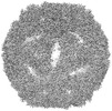 | |||||||||
|---|---|---|---|---|---|---|---|---|---|---|
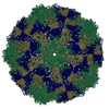
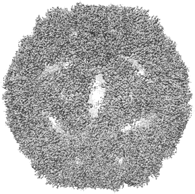
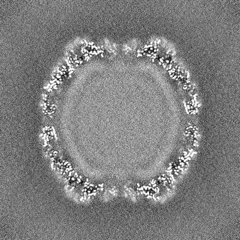
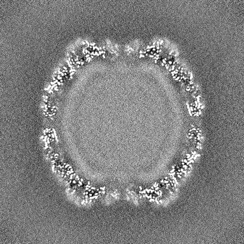
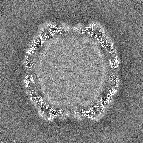
| Title | Structure of coxsackievirus B5 capsid (mutant CVB5F.cas.genogroupB) - A particle | |||||||||
 Map data Map data | Main map | |||||||||
 Sample Sample |
| |||||||||
 Keywords Keywords | Enterovirus / coxsackievirus / thermostable / mutant / VIRUS | |||||||||
| Biological species |  Coxsackievirus B5 Coxsackievirus B5 | |||||||||
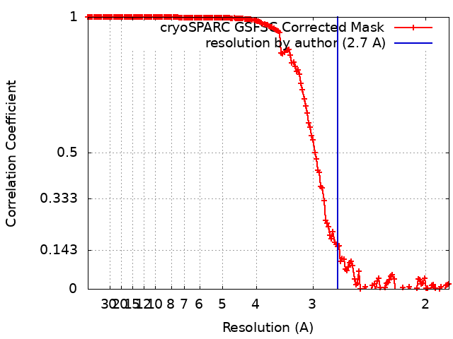
| Method | single particle reconstruction / cryo EM / Resolution: 2.7 Å | |||||||||
 Authors Authors | Kumar K / Antanasijevic A | |||||||||
| Funding support | 1 items
| |||||||||
 Citation Citation | Journal: Environ Sci Technol / Year: 2024 Title: Influence of Amino Acid Substitutions in Capsid Proteins of Coxsackievirus B5 on Free Chlorine and Thermal Inactivation. Authors: Shotaro Torii / Jérôme Gouttenoire / Kiruthika Kumar / Aleksandar Antanasijevic / Tamar Kohn /  Abstract: The sensitivity of enteroviruses to disinfectants varies among genetically similar variants and coincides with amino acid changes in capsid proteins, although the effect of individual substitutions ...The sensitivity of enteroviruses to disinfectants varies among genetically similar variants and coincides with amino acid changes in capsid proteins, although the effect of individual substitutions remains unknown. Here, we employed reverse genetics to investigate how amino acid substitutions in coxsackievirus B5 (CVB5) capsid proteins affect the virus' sensitivity to free chlorine and heat treatment. Of ten amino acid changes observed in CVB5 variants with free chlorine resistance, none significantly reduced the chlorine sensitivity, indicating a minor role of the capsid composition in chlorine sensitivity of CVB5. Conversely, a subset of these amino acid changes located at the C-terminal region of viral protein 1 led to reduced heat sensitivity. Cryo-electron microscopy revealed that these changes affect the assembly of intermediate viral states (altered and empty particles), suggesting that the mechanism for reduced heat sensitivity could be related to improved molecular packing of CVB5, resulting in greater stability or altered dynamics of virus uncoating during infection. | |||||||||
| History |
|
- Structure visualization
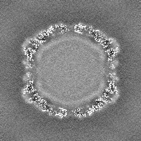
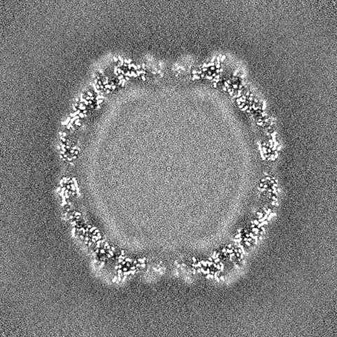
| Supplemental images |
|---|
- Downloads & links
-EMDB archive
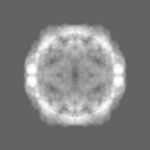
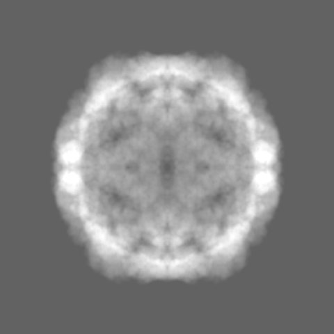
| Map data | emd_18943.map.gz | 398.8 MB |  EMDB map data format EMDB map data format | |
|---|---|---|---|---|
| Header (meta data) | emd-18943-v30.xmlemd-18943.xml | 18.7 KB 18.7 KB | Display Display | EMDB header |
| FSC (resolution estimation) | emd_18943_fsc.xml | 15.8 KB | Display | FSC data file |
| Images |  emd_18943.png emd_18943.png | 237.7 KB | ||
| Masks | emd_18943_msk_1.map | 421.9 MB | Mask map | |
| Filedesc metadata | emd-18943.cif.gz | 6.7 KB | ||
| Others | emd_18943_half_map_1.map.gzemd_18943_half_map_2.map.gz | 391.9 MB 391.9 MB | ||
| Archive directory |  http://ftp.pdbj.org/pub/emdb/structures/EMD-18943ftp://ftp.pdbj.org/pub/emdb/structures/EMD-18943 http://ftp.pdbj.org/pub/emdb/structures/EMD-18943ftp://ftp.pdbj.org/pub/emdb/structures/EMD-18943 | HTTPS FTP |
-Validation report
| Summary document | emd_18943_validation.pdf.gz | 1.3 MB | Display | EMDB validaton report |
|---|---|---|---|---|
| Full document | emd_18943_full_validation.pdf.gz | 1.3 MB | Display | |
| Data in XML | emd_18943_validation.xml.gz | 25 KB | Display | |
| Data in CIF | emd_18943_validation.cif.gz | 32.2 KB | Display | |
| Arichive directory | https://ftp.pdbj.org/pub/emdb/validation_reports/EMD-18943ftp://ftp.pdbj.org/pub/emdb/validation_reports/EMD-18943 | HTTPS FTP |
-Related structure data
| Related structure data | 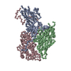 8r5yMC 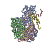 8r5xC  8r5zC C: citing same article ( M: atomic model generated by this map |
|---|

-Links
| EMDB pages | EMDB (EBI/PDBe) / EMDataResource |
|---|
-Map
| File | Download / File: emd_18943.map.gz / Format: CCP4 / Size: 421.9 MB / Type: IMAGE STORED AS FLOATING POINT NUMBER (4 BYTES) | ||||||||||||||||||||
|---|---|---|---|---|---|---|---|---|---|---|---|---|---|---|---|---|---|---|---|---|---|
| Annotation | Main map | ||||||||||||||||||||
| Voxel size | X=Y=Z: 0.926 Å | ||||||||||||||||||||
| Density |
| ||||||||||||||||||||
| Symmetry | Space group: 1 | ||||||||||||||||||||
| Details | EMDB XML:
|
-Supplemental data
-Mask #1
| File | emd_18943_msk_1.map | ||||||||||||
|---|---|---|---|---|---|---|---|---|---|---|---|---|---|
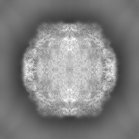
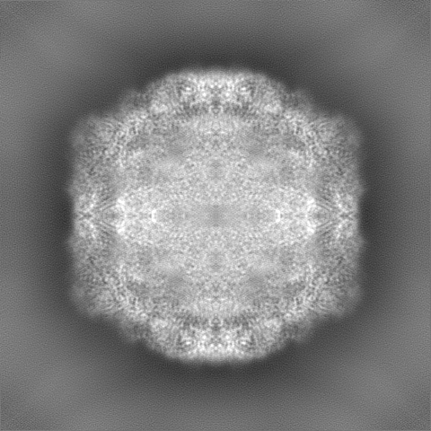
| Projections & Slices |
| ||||||||||||
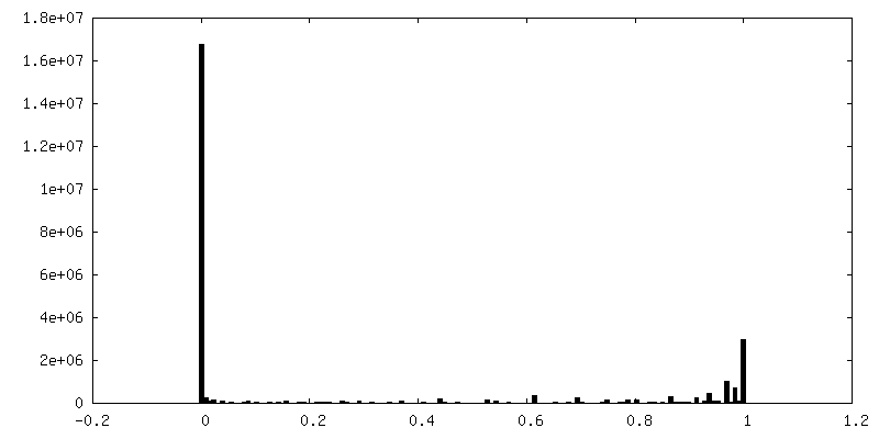
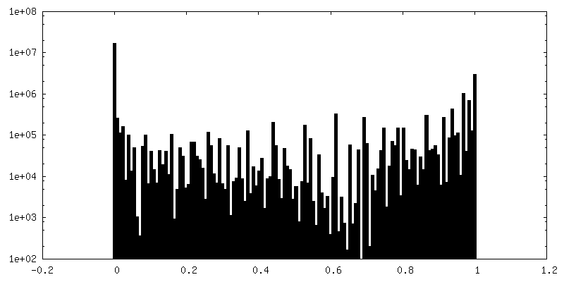
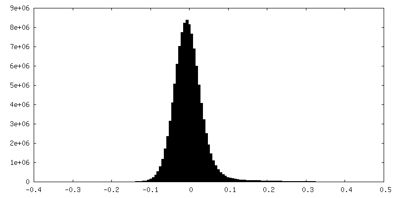
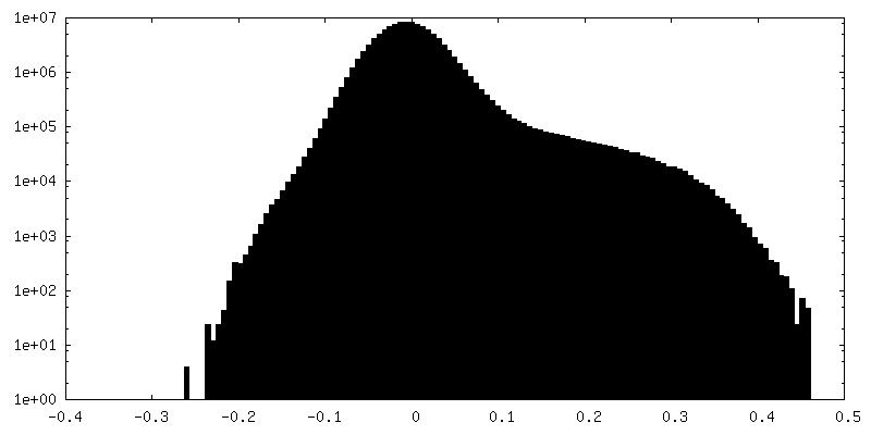
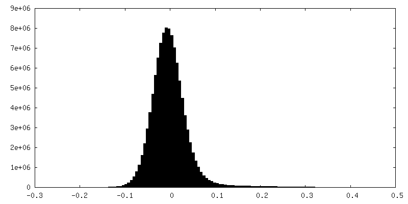
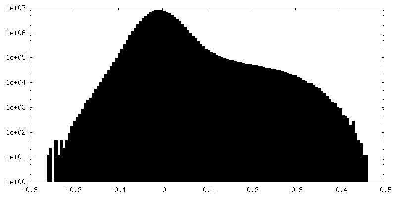
| Density Histograms |
 Z
Z Y
Y X
X


-Half map: Half-Map A
| File | emd_18943_half_map_1.map | ||||||||||||
|---|---|---|---|---|---|---|---|---|---|---|---|---|---|
| Annotation | Half-Map A | ||||||||||||
| Projections & Slices |
| ||||||||||||
| Density Histograms |


-Half map: Half-Map B
| File | emd_18943_half_map_2.map | ||||||||||||
|---|---|---|---|---|---|---|---|---|---|---|---|---|---|
| Annotation | Half-Map B | ||||||||||||
| Projections & Slices |
| ||||||||||||
| Density Histograms |


- Sample components
-Entire : Coxsackievirus B5
| Entire | Name: Coxsackievirus B5 |
|---|---|
| Components |
|
-Supramolecule #1: Coxsackievirus B5
| Supramolecule | Name: Coxsackievirus B5 / type: virus / ID: 1 / Parent: 0 / Macromolecule list: all / Details: Particle A state of the CVB5 virion capsid / NCBI-ID: 12074 / Sci species name: Coxsackievirus B5 / Virus type: VIRION / Virus isolate: STRAIN / Virus enveloped: No / Virus empty: No |
|---|---|
| Host (natural) | Organism:  Homo sapiens (human) Homo sapiens (human) |
| Virus shell | Shell ID: 1 / Name: Capsid VP1-4 / Diameter: 300.0 Å |
-Macromolecule #1: Coxsackievirus B5 (mutant CVB5F.cas.genogroupB) in particle A sta...
| Macromolecule | Name: Coxsackievirus B5 (mutant CVB5F.cas.genogroupB) in particle A state - VP1 type: protein_or_peptide / ID: 1 / Number of copies: 3 / Enantiomer: LEVO |
|---|---|
| Source (natural) | Organism: Coxsackievirus B5 |
| Molecular weight | Theoretical: 93.795359 KDa |
| Recombinant expression | Organism: Homo sapiens (human) |
| Sequence | String: MGAQVSTQKT GAHETGLNAS GNSIIHYTNM NYYKDSASNS ANRQEFAQDP GKFTEPVKDI MIKSMPALNS PSAEECGYSD RVRSITLGN STITTQECAN VVVGYGTWPT YLRDEEATAE DQPTQPDVAT CRFYTLESVM WQQSSPGWWW KFPDALSNMG L FGQNMQYH ...String: MGAQVSTQKT GAHETGLNAS GNSIIHYTNM NYYKDSASNS ANRQEFAQDP GKFTEPVKDI MIKSMPALNS PSAEECGYSD RVRSITLGN STITTQECAN VVVGYGTWPT YLRDEEATAE DQPTQPDVAT CRFYTLESVM WQQSSPGWWW KFPDALSNMG L FGQNMQYH YLGRAGYTLH VQCNASKFHQ GCLLVVCVPE AEMGCATIAN KPDQKSLSNG ETANVFDSQN TSGQTAVQAN VI NAGMGIG VGNLTIFPHQ WINLRTNNSA TIVMPYVNSV PMDNMFRHNN FTLMIIPFAP LSYSTGATTY VPITVTVAPM CAE YNGLRL AGRQGLPTML TPGSNQFLTS DDFQSPSAMP QFDVTPEMAI PGQVNNLMEI AEVDSVVPVN NTEGKVMSIE AYQI PVQSN STNGSQVFGF PLIPGASSVL NRTLLGEILN YYTHWSGSIK LTFMFCGSAM ATGKFLLAYS PPGAGAPTTR KEAML GTHV IWDVGLQSSC VLCIPWISQT HYRYVVMDEY TAGGYITCWY QTNIVVPADT QSDCKILCFV SACNDFSVRM LKDTPF IKQ DSFFQGPPGE AIERAIARVA DTISSGPVNS ESIPALTAAE TGHTSQVVPA DTMQTRHVKN YHSRSESTVE NFLCRSA CV YYTTYKNHGT DGDNFAQWVI NTRQVAQLRR KLEMFTYARF DLELTFVITS SQEQSTIKGQ DSPVLTHQIM YVPPGGPV P TKINSYSWQT STNPSVFWTE GSAPPRISIP FISIGNAYSM FYDGWARFDK QGTYGINTLN NMGTLYMRHV NDGSPGPIV STVRIYFKPK HVKTWVPRPP RLCQYQKAGN VNFEPSGVTE GRTEITAMQT T |
-Experimental details
-Structure determination
| Method | cryo EM |
|---|---|
 Processing Processing | single particle reconstruction |
| Aggregation state | particle |
-Sample preparation
| Concentration | 4.5 mg/mL | |||||||||
|---|---|---|---|---|---|---|---|---|---|---|
| Buffer | pH: 7.5 Component:
Details: TBS | |||||||||
| Vitrification | Cryogen name: ETHANE / Chamber humidity: 100 % / Chamber temperature: 283 K / Instrument: FEI VITROBOT MARK IV Details: 3ul of sample applied. Blotting time varied. Blotting force = 0. Total blots = 1.. | |||||||||
| Details | Inactivated by formaldehyde. Purified using a combination of sucrose gradient and size-exclusion chromatography. |
- Electron microscopy
| Microscope | TFS GLACIOS |
|---|---|
| Image recording | Film or detector model: FEI FALCON IV (4k x 4k) / Number grids imaged: 2 / Number real images: 6653 / Average electron dose: 40.0 e/Å2 |
| Electron beam | Acceleration voltage: 200 kV / Electron source:  FIELD EMISSION GUN FIELD EMISSION GUN |
| Electron optics | Illumination mode: FLOOD BEAM / Imaging mode: BRIGHT FIELD / Cs: 2.7 mm / Nominal defocus max: 1.0 µm / Nominal defocus min: 0.8 µm / Nominal magnification: 150000 |
| Sample stage | Specimen holder model: OTHER / Cooling holder cryogen: NITROGEN |
+Image processing
-Atomic model buiding 1
| Initial model | PDB ID: Chain - Source name: PDB / Chain - Initial model type: experimental model Details: Asymmetric unit used for fitting and interpretation |
|---|---|
| Details | Initial fitting was performed in Chimera and model refinement was performed in Coot and Rosetta. |
| Refinement | Space: REAL / Protocol: RIGID BODY FIT |
| Output model | PDB-8r5y: |