 Movie
Movie Controller

[English] 日本語
 Yorodumi
- PDB-7xb2: CVB5-intermediate altered particle containing VP1/VP2/VP3 and RNA... -
+ Open data

- Basic information
| Entry | Database: PDB / ID: 7xb2 | |||||||||||||||||||||||||||
|---|---|---|---|---|---|---|---|---|---|---|---|---|---|---|---|---|---|---|---|---|---|---|---|---|---|---|---|---|
| Title | CVB5-intermediate altered particle containing VP1/VP2/VP3 and RNA genome | |||||||||||||||||||||||||||
 Components Components | (Genome polyprotein) x 3 | |||||||||||||||||||||||||||
 Keywords Keywords | VIRUS / Coxsackie virus B5 / intermediate altered particle | |||||||||||||||||||||||||||
| Function / homology |  Function and homology information Function and homology informationsymbiont-mediated suppression of host cytoplasmic pattern recognition receptor signaling pathway via inhibition of RIG-I activity / picornain 2A / symbiont-mediated suppression of host mRNA export from nucleus / symbiont genome entry into host cell via pore formation in plasma membrane / picornain 3C / T=pseudo3 icosahedral viral capsid / host cell cytoplasmic vesicle membrane / viral capsid / ribonucleoside triphosphate phosphatase activity / host cell ...symbiont-mediated suppression of host cytoplasmic pattern recognition receptor signaling pathway via inhibition of RIG-I activity / picornain 2A / symbiont-mediated suppression of host mRNA export from nucleus / symbiont genome entry into host cell via pore formation in plasma membrane / picornain 3C / T=pseudo3 icosahedral viral capsid / host cell cytoplasmic vesicle membrane / viral capsid / ribonucleoside triphosphate phosphatase activity / host cell / nucleoside-triphosphate phosphatase / channel activity / monoatomic ion transmembrane transport / DNA replication / RNA helicase activity / endocytosis involved in viral entry into host cell / symbiont-mediated activation of host autophagy / RNA-directed RNA polymerase / cysteine-type endopeptidase activity / viral RNA genome replication / RNA-directed RNA polymerase activity / symbiont entry into host cell / DNA-templated transcription / virion attachment to host cell / host cell nucleus / structural molecule activity / proteolysis / RNA binding / zinc ion binding / ATP binding Similarity search - Function | |||||||||||||||||||||||||||
| Biological species |  Coxsackievirus B5 Coxsackievirus B5 | |||||||||||||||||||||||||||
| Method | ELECTRON MICROSCOPY / single particle reconstruction / cryo EM / Resolution: 2.81 Å | |||||||||||||||||||||||||||
 Authors Authors | Yang, P. / Wang, K. | |||||||||||||||||||||||||||
| Funding support |  China, 1items China, 1items
| |||||||||||||||||||||||||||
 Citation Citation | Journal: J Virol / Year: 2022 Title: Atomic Structures of Coxsackievirus B5 Provide Key Information on Viral Evolution and Survival. Authors: Peng Yang / Dawei Shi / Jianmeng Fu / Li Zhang / Ruihong Chen / Binyang Zheng / Xiangxi Wang / Sihong Xu / Ling Zhu / Kang Wang / Abstract: Coxsackie virus B5 (CVB5), a main serotype in human Enterovirus B (EVB), can cause severe viral encephalitis and aseptic meningitis among infants and children. Currently, there is no approved vaccine ...Coxsackie virus B5 (CVB5), a main serotype in human Enterovirus B (EVB), can cause severe viral encephalitis and aseptic meningitis among infants and children. Currently, there is no approved vaccine or antiviral therapy available against CVB5 infection. Here, we determined the atomic structures of CVB5 in three forms: mature full (F) particle (2.73 Å), intermediate altered (A) particle (2.81 Å), and procapsid empty (E) particle (2.95 Å). Structural analysis of F particle of CVB5 unveiled similar structures of "canyon," "puff," and "knob" as those other EV-Bs. We observed structural rearrangements that are alike during the transition from F to A particle, indicative of similar antigenicity, cell entry, and uncoating mechanisms shared by all EV-Bs. Further comparison of structures and sequences among all structure-known EV-Bs revealed that while the residues targeted by neutralizing MAbs are diversified and drive the evolution of EV-Bs, the relative conserved residues recognized by uncoating receptors could serve as the basis for the development of antiviral vaccines and therapeutics. As one of the main serotypes in Enterovirus B, CVB5 has been commonly reported in recent years. The atomic structures of CVB5 shown here revealed classical features found in EV-Bs and the structural rearrangement occurring during particle expansion and uncoating. Also, structure- and sequence-based comparison between CVB5 and other structure-known EV-Bs screened out key domains important for viral evolution and survival. All these provide insights into the development of vaccine and therapeutics for EV-Bs. | |||||||||||||||||||||||||||
| History |
|
- Structure visualization
| Structure viewer | Molecule: MolmilJmol/JSmol |
|---|
- Downloads & links
-Download
| PDBx/mmCIF format | 7xb2.cif.gz | 137.2 KB | Display | PDBx/mmCIF format |
|---|---|---|---|---|
| PDB format | pdb7xb2.ent.gz | 104.6 KB | Display | PDB format |
| PDBx/mmJSON format | 7xb2.json.gz | Tree view | PDBx/mmJSON format | |
| Others |  Other downloads Other downloads |
-Validation report
| Arichive directory | https://data.pdbj.org/pub/pdb/validation_reports/xb/7xb2ftp://data.pdbj.org/pub/pdb/validation_reports/xb/7xb2 | HTTPS FTP |
|---|
-Related structure data
| Related structure data | 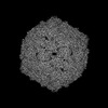 33101MC 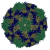 7wl3C M: map data used to model this data C: citing same article ( |
|---|---|
| Similar structure data |
-Links
 PDBj
PDBj

- Assembly
| Deposited unit | 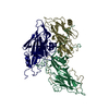
|
|---|---|
| 1 | x 60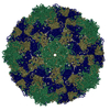
|
| 2 |
|
| 3 | x 5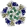
|
| 4 | x 6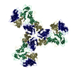
|
| 5 | 
|
| Symmetry | Point symmetry: (Schoenflies symbol: I (icosahedral)) |
-Components
| #1: Protein | Mass: 26653.973 Da / Num. of mol.: 1 Source method: isolated from a genetically manipulated source Source: (gene. exp.) Coxsackievirus B5 / Gene: VP1 / Production host:  Homo sapiens (human) / References: UniProt: A0A348FI90 Homo sapiens (human) / References: UniProt: A0A348FI90 |
|---|---|
| #2: Protein | Mass: 27242.725 Da / Num. of mol.: 1 Source method: isolated from a genetically manipulated source Source: (gene. exp.) Coxsackievirus B5 / Gene: polyprotein / Production host: Homo sapiens (human)References: UniProt: A0A6M4MJ36, picornain 2A, nucleoside-triphosphate phosphatase, picornain 3C, RNA-directed RNA polymerase |
| #3: Protein | Mass: 26163.672 Da / Num. of mol.: 1 Source method: isolated from a genetically manipulated source Source: (gene. exp.) Coxsackievirus B5 / Gene: polyprotein / Production host: Homo sapiens (human)References: UniProt: A0A6M4MJ36, picornain 2A, nucleoside-triphosphate phosphatase, picornain 3C, RNA-directed RNA polymerase |
| Has protein modification | N |
-Experimental details
-Experiment
| Experiment | Method: ELECTRON MICROSCOPY |
|---|---|
| EM experiment | Aggregation state: PARTICLE / 3D reconstruction method: single particle reconstruction |
- Sample preparation
| Component | Name: Coxsackievirus B5 / Type: VIRUS / Details: Coxsackievirus B5-A-particle / Entity ID: all / Source: NATURAL |
|---|---|
| Source (natural) | Organism: Coxsackievirus B5 |
| Details of virus | Empty: NO / Enveloped: NO / Isolate: STRAIN / Type: VIRION |
| Buffer solution | pH: 7.4 / Details: PBS buffer |
| Specimen | Conc.: 1.5 mg/ml / Embedding applied: NO / Shadowing applied: NO / Staining applied: NO / Vitrification applied: YES |
| Vitrification | Cryogen name: ETHANE / Humidity: 100 % / Chamber temperature: 25 K |
- Electron microscopy imaging
| Experimental equipment |  Model: Titan Krios / Image courtesy: FEI Company |
|---|---|
| Microscopy | Model: FEI TITAN KRIOS |
| Electron gun | Electron source:  FIELD EMISSION GUN / Accelerating voltage: 300 kV / Illumination mode: OTHER FIELD EMISSION GUN / Accelerating voltage: 300 kV / Illumination mode: OTHER |
| Electron lens | Mode: DIFFRACTION / Nominal defocus max: 2500 nm / Nominal defocus min: 1200 nm |
| Image recording | Electron dose: 30 e/Å2 / Film or detector model: GATAN K2 BASE (4k x 4k) |
- Processing
| Software | Name: PHENIX / Version: 1.19.2_4158: / Classification: refinement | ||||||||||||||||||||||||
|---|---|---|---|---|---|---|---|---|---|---|---|---|---|---|---|---|---|---|---|---|---|---|---|---|---|
| EM software | Name: PHENIX / Category: model refinement | ||||||||||||||||||||||||
| CTF correction | Type: PHASE FLIPPING ONLY | ||||||||||||||||||||||||
| 3D reconstruction | Resolution: 2.81 Å / Resolution method: FSC 0.143 CUT-OFF / Num. of particles: 7675 / Symmetry type: POINT | ||||||||||||||||||||||||
| Refine LS restraints |
|
