ムービー
ムービー コントローラー


+ データを開く

- 基本情報
基本情報
| 登録情報 | データベース: EMDB / ID: EMD-1713 | |||||||||
|---|---|---|---|---|---|---|---|---|---|---|
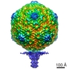
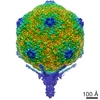
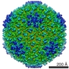
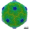
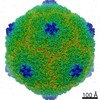
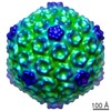
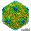
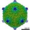
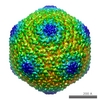
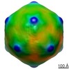
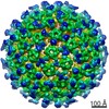
| タイトル | Capsid structure of the infectious Prochlorococcus Cyanophage P-SSP7 | |||||||||
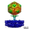
 マップデータ マップデータ | Icosahedral capsid structure of cyanophage P-SSP7 | |||||||||
 試料 試料 |
| |||||||||
 キーワード キーワード | Marine Podovirus / T7-like virus / infects Prochlorococcus MED4 | |||||||||
| 機能・相同性 | : / Major capsid protein / T7-like capsid protein 機能・相同性情報 機能・相同性情報 | |||||||||
| 生物種 |  Prochlorococcus phage P-SSP7 (ファージ) Prochlorococcus phage P-SSP7 (ファージ) | |||||||||
| 手法 | 単粒子再構成法 / クライオ電子顕微鏡法 / ネガティブ染色法 / 解像度: 4.6 Å | |||||||||
 データ登録者 データ登録者 | Liu X / Zhang Q / Murata K / Baker ML / Sullivan MB / Fu C / Dougherty M / Schmid MF / Osburne MS / Chisholm SW / Chiu W | |||||||||
 引用 引用 | ジャーナル: Nat Struct Mol Biol / 年: 2010 タイトル: Structural changes in a marine podovirus associated with release of its genome into Prochlorococcus. 著者: Xiangan Liu / Qinfen Zhang / Kazuyoshi Murata / Matthew L Baker / Matthew B Sullivan / Caroline Fu / Matthew T Dougherty / Michael F Schmid / Marcia S Osburne / Sallie W Chisholm / Wah Chiu /  要旨: Podovirus P-SSP7 infects Prochlorococcus marinus, the most abundant oceanic photosynthetic microorganism. Single-particle cryo-electron microscopy yields icosahedral and asymmetrical structures of ...Podovirus P-SSP7 infects Prochlorococcus marinus, the most abundant oceanic photosynthetic microorganism. Single-particle cryo-electron microscopy yields icosahedral and asymmetrical structures of infectious P-SSP7 with 4.6-A and 9-A resolution, respectively. The asymmetric reconstruction reveals how symmetry mismatches are accommodated among five of the gene products at the portal vertex. Reconstructions of infectious and empty particles show a conformational change of the 'valve' density in the nozzle, an orientation difference in the tail fibers, a disordering of the C terminus of the portal protein and the disappearance of the core proteins. In addition, cryo-electron tomography of P-SSP7 infecting Prochlorococcus showed the same tail-fiber conformation as that in empty particles. Our observations suggest a mechanism whereby, upon binding to the host cell, the tail fibers induce a cascade of structural alterations of the portal vertex complex that triggers DNA release. | |||||||||
| 履歴 |
|
- 構造の表示
構造の表示
| ムービー |
ムービービューア |
|---|---|
| 構造ビューア | EMマップ: SurfViewMolmilJmol/JSmol |
| 添付画像 |
- ダウンロードとリンク
-EMDBアーカイブ
| マップデータ | emd_1713.map.gz | 97.9 MB | EMDBマップデータ形式 | |
|---|---|---|---|---|
| ヘッダ (付随情報) | emd-1713-v30.xmlemd-1713.xml | 12.1 KB 12.1 KB | 表示 表示 | EMDBヘッダ |
| 画像 | 412.9 KB | |||
| アーカイブディレクトリ |  http://ftp.pdbj.org/pub/emdb/structures/EMD-1713ftp://ftp.pdbj.org/pub/emdb/structures/EMD-1713 http://ftp.pdbj.org/pub/emdb/structures/EMD-1713ftp://ftp.pdbj.org/pub/emdb/structures/EMD-1713 | HTTPS FTP |
-関連構造データ

-リンク
| EMDBのページ | EMDB (EBI/PDBe) / EMDataResource |
|---|
-マップ
| ファイル | ダウンロード / ファイル: emd_1713.map.gz / 形式: CCP4 / 大きさ: 711.9 MB / タイプ: IMAGE STORED AS FLOATING POINT NUMBER (4 BYTES) | ||||||||||||||||||||||||||||||||||||||||||||||||||||||||||||||||||||
|---|---|---|---|---|---|---|---|---|---|---|---|---|---|---|---|---|---|---|---|---|---|---|---|---|---|---|---|---|---|---|---|---|---|---|---|---|---|---|---|---|---|---|---|---|---|---|---|---|---|---|---|---|---|---|---|---|---|---|---|---|---|---|---|---|---|---|---|---|---|
| 注釈 | Icosahedral capsid structure of cyanophage P-SSP7 | ||||||||||||||||||||||||||||||||||||||||||||||||||||||||||||||||||||
| 投影像・断面図 | 画像のコントロール
画像は Spider により作成 | ||||||||||||||||||||||||||||||||||||||||||||||||||||||||||||||||||||
| ボクセルのサイズ | X=Y=Z: 1.17 Å | ||||||||||||||||||||||||||||||||||||||||||||||||||||||||||||||||||||
| 密度 |
| ||||||||||||||||||||||||||||||||||||||||||||||||||||||||||||||||||||
| 対称性 | 空間群: 1 | ||||||||||||||||||||||||||||||||||||||||||||||||||||||||||||||||||||
| 詳細 | EMDB XML:
CCP4マップ ヘッダ情報:
| ||||||||||||||||||||||||||||||||||||||||||||||||||||||||||||||||||||
 Z (Sec.)
 Y (Row.)
 X (Col.)


-添付データ
- 試料の構成要素
-全体 : Cyanophage P-SSP7
| 全体 | 名称: Cyanophage P-SSP7 |
|---|---|
| 要素 |
|
-超分子 #1000: Cyanophage P-SSP7
| 超分子 | 名称: Cyanophage P-SSP7 / タイプ: sample / ID: 1000 / Number unique components: 1 |
|---|
-超分子 #1: Prochlorococcus phage P-SSP7
| 超分子 | 名称: Prochlorococcus phage P-SSP7 / タイプ: virus / ID: 1 / Name.synonym: T7-LIKE CAPSID 詳細: The gp10 protein of P-SSP7 contains 375 amino acids. NCBI-ID: 268748 / 生物種: Prochlorococcus phage P-SSP7 / ウイルスタイプ: VIRION / ウイルス・単離状態: STRAIN / ウイルス・エンベロープ: No / ウイルス・中空状態: No / Syn species name: T7-LIKE CAPSID |
|---|---|
| 宿主 | 生物種:  Prochlorococcus (バクテリア) / 別称: BACTERIA(EUBACTERIA) Prochlorococcus (バクテリア) / 別称: BACTERIA(EUBACTERIA) |
| ウイルス殻 | Shell ID: 1 / 直径: 655 Å / T番号(三角分割数): 7 |
-実験情報
-構造解析
| 手法 | ネガティブ染色法, クライオ電子顕微鏡法 |
|---|---|
 解析 解析 | 単粒子再構成法 |
| 試料の集合状態 | particle |
-試料調製
| 濃度 | 5 mg/mL |
|---|---|
| 緩衝液 | pH: 7.5 / 詳細: 100 mM Tris-HCl , 100 mM MgSO4, and 30 mM NaCl |
| 染色 | タイプ: NEGATIVE / 詳細: No stain |
| グリッド | 詳細: 200 mesh copper grid |
| 凍結 | 凍結剤: ETHANE / チャンバー内湿度: 30 % / チャンバー内温度: 101 K / 装置: OTHER / 詳細: Vitrification instrument: FEI Vitrobot / 手法: Blot for 2 seconds 2 times before plunging |
- 電子顕微鏡法
| 顕微鏡 | JEOL 3200FSC |
|---|---|
| 温度 | 最低: 4.5 K / 最高: 102 K / 平均: 101 K |
| アライメント法 | Legacy - 非点収差: Objective lens astigmatism was corrected at 400,000 times magnification |
| 特殊光学系 | エネルギーフィルター - 名称: JEOL エネルギーフィルター - エネルギー下限: 15.0 eV エネルギーフィルター - エネルギー上限: 20.0 eV |
| 詳細 | MDS |
| 日付 | 2007年8月31日 |
| 撮影 | カテゴリ: FILM / フィルム・検出器のモデル: KODAK SO-163 FILM デジタル化 - スキャナー: NIKON SUPER COOLSCAN 9000 デジタル化 - サンプリング間隔: 6.35 µm / 実像数: 1059 / 平均電子線量: 20 e/Å2 詳細: The cryoEM images were recorded on Kodak SO163 films. The films were developed in full strength D19 Kodak developer for 12 minutes at 20 degree and fixed for 10 minutes in Kodak fixer. The ...詳細: The cryoEM images were recorded on Kodak SO163 films. The films were developed in full strength D19 Kodak developer for 12 minutes at 20 degree and fixed for 10 minutes in Kodak fixer. The films were digitized at 6.35 microns per pixel using a Nikon Super CoolScan 9000 ED scanner (Nikon Corp., Japan). ビット/ピクセル: 8 |
| 電子線 | 加速電圧: 300 kV / 電子線源:  FIELD EMISSION GUN FIELD EMISSION GUN |
| 電子光学系 | 照射モード: SPOT SCAN / 撮影モード: BRIGHT FIELD / Cs: 4.1 mm / 最大 デフォーカス(公称値): 3.0 µm / 最小 デフォーカス(公称値): 0.6 µm / 倍率(公称値): 60000 |
| 試料ステージ | 試料ホルダー: Side entry / 試料ホルダーモデル: JEOL 3200FSC CRYOHOLDER |
-画像解析
| 詳細 | The particle were selected by the consistency criterion of MPSA |
|---|---|
| CTF補正 | 詳細: Each Micrograph |
| 最終 再構成 | 想定した対称性 - 点群: I (正20面体型対称) / アルゴリズム: OTHER / 解像度のタイプ: BY AUTHOR / 解像度: 4.6 Å / 解像度の算出法: FSC 0.5 CUT-OFF / ソフトウェア - 名称: MPSA 詳細: The map was directly built from raw particles with icosahedral symmetry enforced. 使用した粒子像数: 36000 |
| 最終 角度割当 | 詳細: EMAN: Z along 5-fold and Y along 2-fold axis. |
-原子モデル構築 1
| 初期モデル | PDB ID: |
|---|---|
| 精密化 | 空間: REAL |
| 得られたモデル | 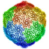 PDB-2xd8: |