 ムービー
ムービー コントローラー


+ データを開く

- 基本情報
基本情報
| 登録情報 |  | |||||||||
|---|---|---|---|---|---|---|---|---|---|---|
| タイトル | Complex III2 from Yarrowia lipolytica, oxidised with ferricyanide, int-position | |||||||||
 マップデータ マップデータ | ||||||||||
 試料 試料 |
| |||||||||
| 機能・相同性 |  機能・相同性情報 機能・相同性情報 mitochondrial processing peptidase complex / mitochondrial processing peptidase / matrix side of mitochondrial inner membrane / protein processing involved in protein targeting to mitochondrion / mitochondrial respiratory chain complex III assembly / mitochondrial respiratory chain complex III / quinol-cytochrome-c reductase / ubiquinol-cytochrome-c reductase activity / mitochondrial electron transport, ubiquinol to cytochrome c / mitochondrial crista ...mitochondrial processing peptidase complex / mitochondrial processing peptidase / matrix side of mitochondrial inner membrane / protein processing involved in protein targeting to mitochondrion / mitochondrial respiratory chain complex III assembly / mitochondrial respiratory chain complex III / quinol-cytochrome-c reductase / ubiquinol-cytochrome-c reductase activity / mitochondrial electron transport, ubiquinol to cytochrome c / mitochondrial crista / nuclear periphery / ミトコンドリア / 2 iron, 2 sulfur cluster binding / metalloendopeptidase activity / oxidoreductase activity / heme binding / ミトコンドリア / タンパク質分解 / metal ion binding / 細胞膜 mitochondrial processing peptidase complex / mitochondrial processing peptidase / matrix side of mitochondrial inner membrane / protein processing involved in protein targeting to mitochondrion / mitochondrial respiratory chain complex III assembly / mitochondrial respiratory chain complex III / quinol-cytochrome-c reductase / ubiquinol-cytochrome-c reductase activity / mitochondrial electron transport, ubiquinol to cytochrome c / mitochondrial crista ...mitochondrial processing peptidase complex / mitochondrial processing peptidase / matrix side of mitochondrial inner membrane / protein processing involved in protein targeting to mitochondrion / mitochondrial respiratory chain complex III assembly / mitochondrial respiratory chain complex III / quinol-cytochrome-c reductase / ubiquinol-cytochrome-c reductase activity / mitochondrial electron transport, ubiquinol to cytochrome c / mitochondrial crista / nuclear periphery / ミトコンドリア / 2 iron, 2 sulfur cluster binding / metalloendopeptidase activity / oxidoreductase activity / heme binding / ミトコンドリア / タンパク質分解 / metal ion binding / 細胞膜類似検索 - 分子機能 | |||||||||
| 生物種 |  Yarrowia lipolytica (酵母) Yarrowia lipolytica (酵母) | |||||||||
| 手法 | 単粒子再構成法 / クライオ電子顕微鏡法 / 解像度: 2.3 Å | |||||||||
 データ登録者 データ登録者 | Wieferig JP / Kuhlbrandt W | |||||||||
| 資金援助 |  ドイツ, 1件 ドイツ, 1件
| |||||||||
 引用 引用 | ジャーナル: IUCrJ / 年: 2023 タイトル: Analysis of the conformational heterogeneity of the Rieske iron-sulfur protein in complex III by cryo-EM. 著者: Jan Philip Wieferig / Werner Kühlbrandt / 要旨: Movement of the Rieske domain of the iron-sulfur protein is essential for intramolecular electron transfer within complex III (CIII) of the respiratory chain as it bridges a gap in the cofactor chain ...Movement of the Rieske domain of the iron-sulfur protein is essential for intramolecular electron transfer within complex III (CIII) of the respiratory chain as it bridges a gap in the cofactor chain towards the electron acceptor cytochrome c. We present cryo-EM structures of CIII from Yarrowia lipolytica at resolutions up to 2.0 Å under different conditions, with different redox states of the cofactors of the high-potential chain. All possible permutations of three primary positions were observed, indicating that the two halves of the dimeric complex act independently. Addition of the substrate analogue decylubiquinone to CIII with a reduced high-potential chain increased the occupancy of the Q site. The extent of Rieske domain interactions through hydrogen bonds to the cytochrome b and cytochrome c subunits varied depending on the redox state and substrate. In the absence of quinols, the reduced Rieske domain interacted more closely with cytochrome b and cytochrome c than in the oxidized state. Upon addition of the inhibitor antimycin A, the heterogeneity of the cd-helix and ef-loop increased, which may be indicative of a long-range effect on the Rieske domain. | |||||||||
| 履歴 |
|
- 構造の表示
構造の表示
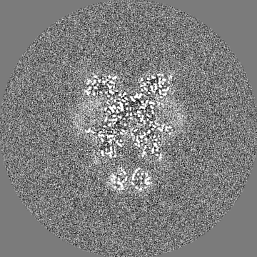
| 添付画像 |
|---|

- ダウンロードとリンク
-EMDBアーカイブ
| マップデータ | emd_15319.map.gz | 165.8 MB | EMDBマップデータ形式 | |
|---|---|---|---|---|
| ヘッダ (付随情報) | emd-15319-v30.xmlemd-15319.xml | 26.2 KB 26.2 KB | 表示 表示 | EMDBヘッダ |
| FSC (解像度算出) | emd_15319_fsc.xml | 12.7 KB | 表示 | FSCデータファイル |
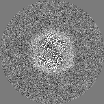
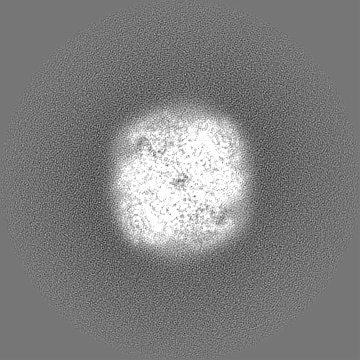
| 画像 |  emd_15319.png emd_15319.png | 61.3 KB | ||
| その他 | emd_15319_half_map_1.map.gzemd_15319_half_map_2.map.gz | 140.9 MB 140.9 MB | ||
| アーカイブディレクトリ |  http://ftp.pdbj.org/pub/emdb/structures/EMD-15319ftp://ftp.pdbj.org/pub/emdb/structures/EMD-15319 http://ftp.pdbj.org/pub/emdb/structures/EMD-15319ftp://ftp.pdbj.org/pub/emdb/structures/EMD-15319 | HTTPS FTP |
-関連構造データ
| 関連構造データ |  8abfMC  8ab6C  8ab7C 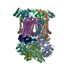 8ab8C  8ab9C  8abaC  8abbC  8abeC  8abgC  8abhC  8abiC  8abjC 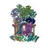 8abkC  8ablC  8abmC  8ac3C  8ac4C  8ac5C M: このマップから作成された原子モデル C: 同じ文献を引用 ( |
|---|---|
| 類似構造データ |


-リンク
| EMDBのページ | EMDB (EBI/PDBe) / EMDataResource |
|---|---|
| 「今月の分子」の関連する項目 |


-マップ
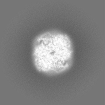
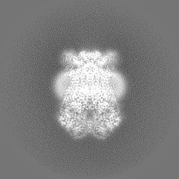
| ファイル | ダウンロード / ファイル: emd_15319.map.gz / 形式: CCP4 / 大きさ: 178 MB / タイプ: IMAGE STORED AS FLOATING POINT NUMBER (4 BYTES) | ||||||||||||||||||||||||||||||||||||
|---|---|---|---|---|---|---|---|---|---|---|---|---|---|---|---|---|---|---|---|---|---|---|---|---|---|---|---|---|---|---|---|---|---|---|---|---|---|
| 投影像・断面図 | 画像のコントロール
画像は Spider により作成 | ||||||||||||||||||||||||||||||||||||
| ボクセルのサイズ | X=Y=Z: 0.837 Å | ||||||||||||||||||||||||||||||||||||
| 密度 |
| ||||||||||||||||||||||||||||||||||||
| 対称性 | 空間群: 1 | ||||||||||||||||||||||||||||||||||||
| 詳細 | EMDB XML:
|
 Z (Sec.)
 Y (Row.)
 X (Col.)


-添付データ
-ハーフマップ: #1
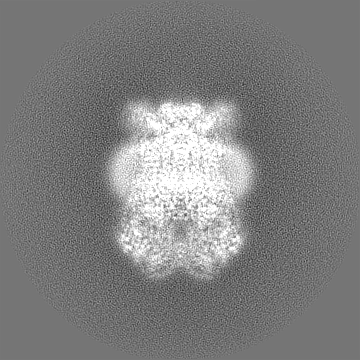
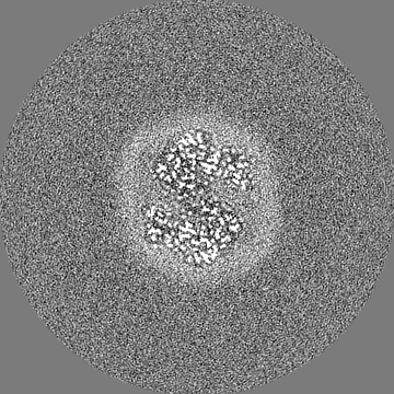
| ファイル | emd_15319_half_map_1.map | ||||||||||||
|---|---|---|---|---|---|---|---|---|---|---|---|---|---|
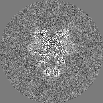
| 投影像・断面図 |
| ||||||||||||
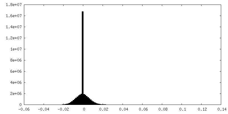
| 密度ヒストグラム |


-ハーフマップ: #2
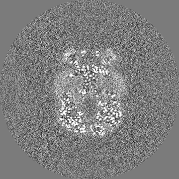
| ファイル | emd_15319_half_map_2.map | ||||||||||||
|---|---|---|---|---|---|---|---|---|---|---|---|---|---|
| 投影像・断面図 |
| ||||||||||||
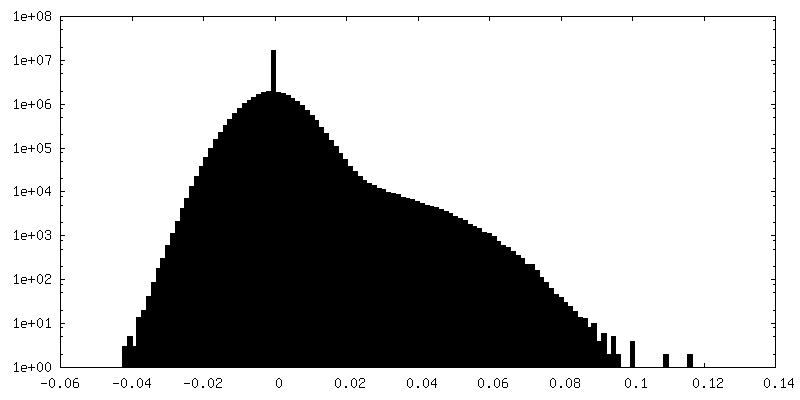
| 密度ヒストグラム |


- 試料の構成要素
+全体 : Complex III2, oxidised with ferricyanide, int-position
+超分子 #1: Complex III2, oxidised with ferricyanide, int-position
+分子 #1: Cytochrome b
+分子 #2: Cytochrome b-c1 complex subunit Rieske, mitochondrial
+分子 #3: Cytochrome b-c1 complex subunit 7
+分子 #4: YALI0F24673p
+分子 #5: YALI0A14806p
+分子 #6: Cytochrome b-c1 complex subunit 2, mitochondrial
+分子 #7: YALI0A17468p
+分子 #8: Cytochrome b-c1 complex subunit 8
+分子 #9: Complex III subunit 9
+分子 #10: YALI0C12210p
+分子 #11: PROTOPORPHYRIN IX CONTAINING FE

+分子 #12: 1,2-DIACYL-SN-GLYCERO-3-PHOSPHOCHOLINE

+分子 #13: PHOSPHATIDYLETHANOLAMINE

+分子 #14: CARDIOLIPIN

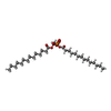
+分子 #15: DODECYL-BETA-D-MALTOSIDE

+分子 #16: FE2/S2 (INORGANIC) CLUSTER

+分子 #17: HEME C

+分子 #18: 1,2-DIMYRISTOYL-SN-GLYCERO-3-PHOSPHATE
-実験情報
-構造解析
| 手法 | クライオ電子顕微鏡法 |
|---|---|
 解析 解析 | 単粒子再構成法 |
| 試料の集合状態 | particle |
-試料調製
| 緩衝液 | pH: 8 |
|---|---|
| グリッド | モデル: UltrAuFoil R1.2/1.3 / 前処理 - タイプ: GLOW DISCHARGE |
| 凍結 | 凍結剤: ETHANE / 装置: FEI VITROBOT MARK IV |
- 電子顕微鏡法
| 顕微鏡 | TFS KRIOS |
|---|---|
| 電子線 | 加速電圧: 300 kV / 電子線源: FIELD EMISSION GUN |
| 電子光学系 | 照射モード: OTHER / 撮影モード: BRIGHT FIELDBright-field microscopy / 最大 デフォーカス(公称値): 2.5 µm / 最小 デフォーカス(公称値): 0.8 µm |
| 試料ステージ | ホルダー冷却材: NITROGEN |
| 撮影 | フィルム・検出器のモデル: GATAN K3 BIOQUANTUM (6k x 4k) 平均電子線量: 55.0 e/Å2 |
| 実験機器 |  モデル: Titan Krios / 画像提供: FEI Company |
-画像解析
| 初期 角度割当 | タイプ: OTHER / ソフトウェア - 名称: RELION |
|---|---|
| 最終 3次元分類 | クラス数: 4 / ソフトウェア - 名称: RELION |
| 最終 角度割当 | タイプ: OTHER / ソフトウェア - 名称: RELION |
| 最終 再構成 | 想定した対称性 - 点群: C1 (非対称) / 解像度のタイプ: BY AUTHOR / 解像度: 2.3 Å / 解像度の算出法: FSC 0.143 CUT-OFF / ソフトウェア - 名称: RELION / 使用した粒子像数: 99452 |
| FSC曲線 (解像度の算出) | 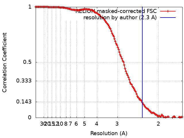 |
